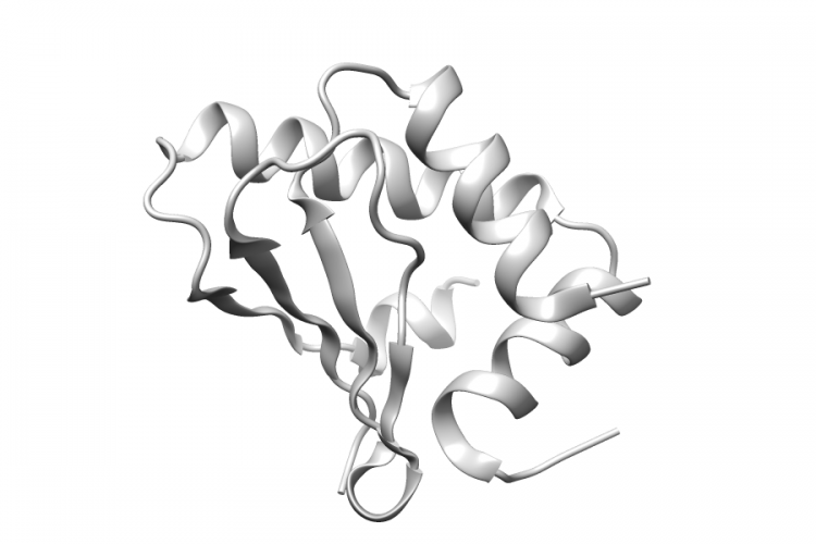
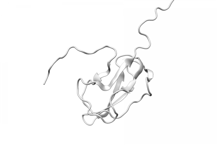
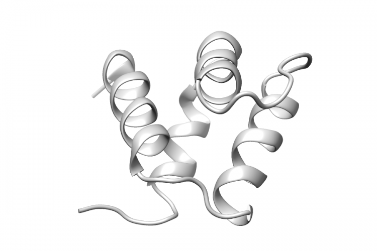
1V66.A | PIAS1

General Structure Information 1v66 PIAS1 solution structure of human p53 binding domain of pias-1 -1.0Å AUTH S.OKUBO,F.HARA,Y.TSUCHIDA,S.SHIMOTAKAHARA,S.SUZUKI,AUTH 2 H.HATANAKA,S.YOKOYAMA,H.TANAKA,H.YASUDA,H.SHINDOTITL NMR STRUCTURE OF THE N-TERMINAL DOMAIN OF SUMOTITL 2 LIGASE PIAS1 AND ITS INTERACTION WITH TUMORTITL 3 SUPPRESSOR P53 AND A/T-RICH DNA OLIGOMERSREF J.BIOL.CHEM. V. 279 31455 2004REFN ISSN 0021-9258PMID 15133049DOI 10.1074/JBC.M403561200 Variant Set Distributions Mapped […]
See Details
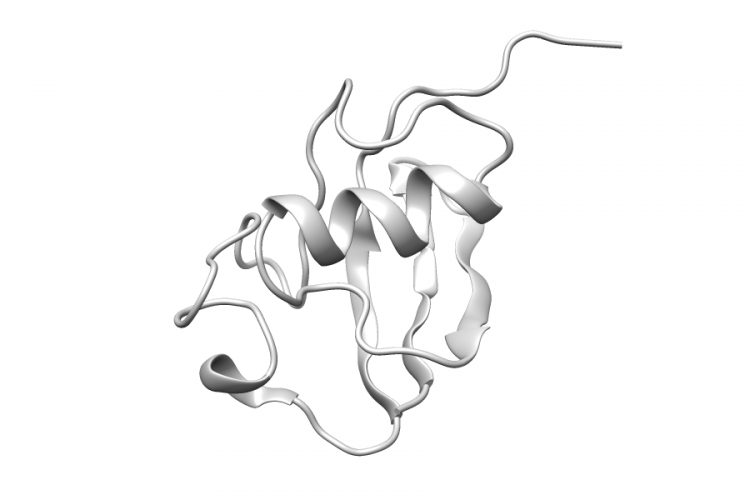
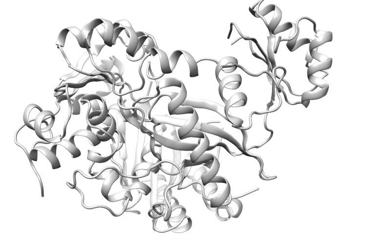
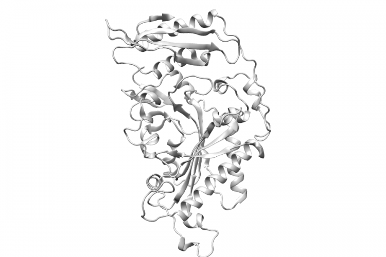
3HFV.A | FARS2

General Structure Information 3hfv FARS2 crystal structure of human mitochondrial phenylalanyl-trna synthetase complexed with m-tyrosine 2.6Å AUTH L.KLIPCAN,N.MOOR,N.KESSLER,M.G.SAFROTITL EUKARYOTIC CYTOSOLIC AND MITOCHONDRIALTITL 2 PHENYLALANYL-TRNA SYNTHETASES CATALYZE THE CHARGINGTITL 3 OF TRNA WITH THE META-TYROSINEREF PROC.NATL.ACAD.SCI.USA V. 106 11045 2009REFN ISSN 0027-8424PMID 19549855DOI 10.1073/PNAS.0905212106 Variant Set Distributions Ripley’s K Analysis Plots Variant Set Comparisons Mapped Variants

See Details
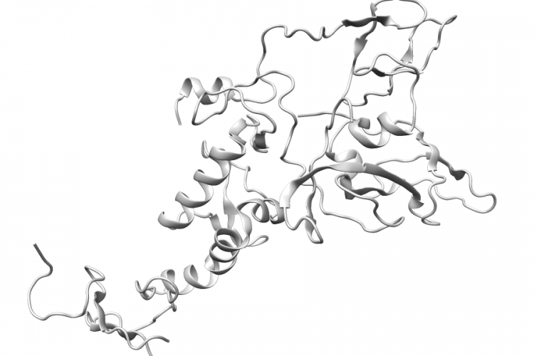
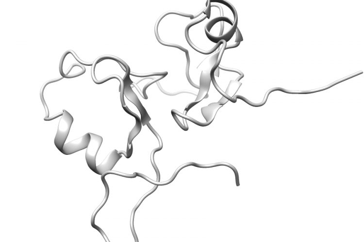
3PT3.B | UBR5

General Structure Information 3pt3 UBR5 crystal structure of the c-terminal lobe of the human ubr5 hect domain 1.97Å AUTH E.MATTA-CAMACHO,G.KOZLOV,M.MENADE,K.GEHRINGTITL STRUCTURE OF THE HECT C-LOBE OF THE UBR5 E3 UBIQUITINTITL 2 LIGASE.REF ACTA CRYSTALLOGR.,SECT.F V. 68 1158 2012REFN ESSN 1744-3091PMID 23027739DOI 10.1107/S1744309112036937 Variant Set Distributions Mapped Variants

See Details
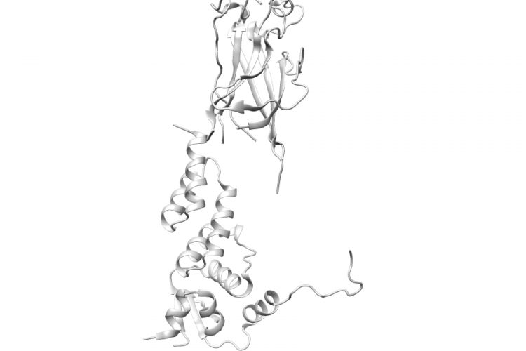
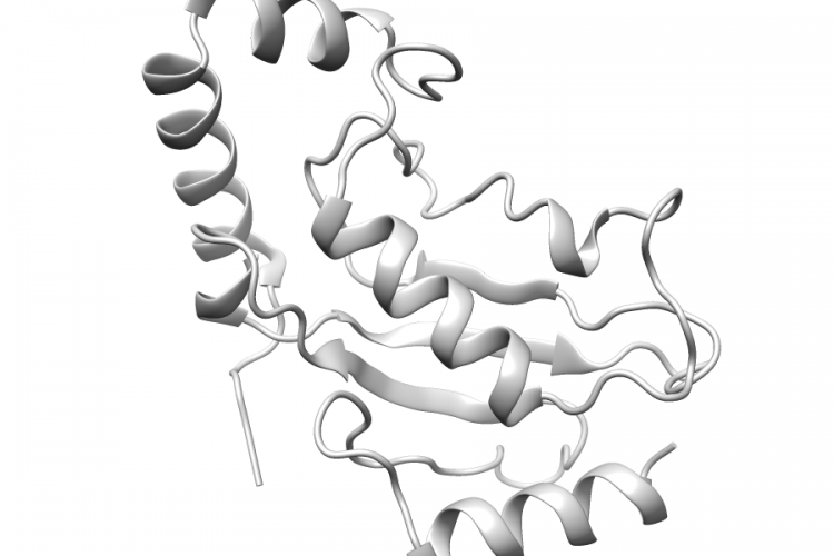
1IYF.A | PARK2

General Structure Information 1iyf PARK2 solution structure of ubiquitin-like domain of human parkin -1.0Å AUTH E.SAKATA,Y.YAMAGUCHI,E.KURIMOTO,J.KIKUCHI,AUTH 2 S.YOKOYAMA,S.YAMADA,H.KAWAHARA,H.YOKOSAWA,AUTH 3 N.HATTORI,Y.MIZUNO,K.TANAKA,K.KATOTITL PARKIN BINDS THE RPN10 SUBUNIT OF 26S PROTEASOMESTITL 2 THROUGH ITS UBIQUITIN-LIKE DOMAINREF EMBO REP. V. 4 301 2003REFN ISSN 1469-221XPMID 12634850DOI 10.1038/SJ.EMBOR.EMBOR764 Variant Set Distributions Mapped Variants

See Details
4I1H.A | PARK2

General Structure Information 4i1h PARK2 structure of parkin e3 ligase 2.0Å AUTH B.E.RILEY,J.C.LOUGHEED,K.CALLAWAY,M.VELASQUEZ,E.BRECHT,AUTH 2 L.NGUYEN,T.SHALER,D.WALKER,Y.YANG,K.REGNSTROM,L.DIEP,AUTH 3 Z.ZHANG,S.CHIOU,M.BOVA,D.R.ARTIS,N.YAO,J.BAKER,T.YEDNOCK,AUTH 4 J.A.JOHNSTONTITL STRUCTURE AND FUNCTION OF PARKIN E3 UBIQUITIN LIGASE REVEALSTITL 2 ASPECTS OF RING AND HECT LIGASES.REF NAT COMMUN V. 4 1982 2013REFN ESSN 2041-1723PMID 23770887DOI 10.1038/NCOMMS2982 Variant Set Distributions Ripley’s K Analysis Plots Variant Set Comparisons […]

See Details
3HU6.A | SPOP

General Structure Information 3hu6 SPOP structures of spop-substrate complexes: insights into molecular architectures of btb-cul3 ubiquitin ligases: spopmathx/btb/3-box-pucsbc1 2.7Å AUTH M.ZHUANG,M.F.CALABRESE,J.LIU,M.B.WADDELL,A.NOURSE,AUTH 2 M.HAMMEL,D.J.MILLER,H.WALDEN,D.M.DUDA,S.N.SEYEDIN,AUTH 3 T.HOGGARD,J.W.HARPER,K.P.WHITE,B.A.SCHULMANTITL STRUCTURES OF SPOP-SUBSTRATE COMPLEXES: INSIGHTSTITL 2 INTO MOLECULAR ARCHITECTURES OF BTB-CUL3 UBIQUITINTITL 3 LIGASES.REF MOL.CELL V. 36 39 2009REFN ISSN 1097-2765PMID 19818708DOI 10.1016/J.MOLCEL.2009.09.022 Variant Set Distributions Ripley’s K Analysis Plots […]

See Details
2DN8.A | ACACB

General Structure Information 2dn8 ACACB solution structure of rsgi ruh-053, an apo-biotin carboxy carrier protein from human transcarboxylase -1.0Å AUTH A.Z.M.RUHUL MOMEN,H.HIROTA,F.HAYASHI,S.YOKOYAMATITL SOLUTION STRUCTURE OF RSGI RUH-053, AN APO-BIOTINTITL 2 CARBOXY CARRIER PROTEIN FROM HUMAN TRANSCARBOXYLASEREF TO BE PUBLISHEDREFN Variant Set Distributions Mapped Variants

See Details
3GID.A | ACACB

General Structure Information 3gid ACACB the biotin carboxylase (bc) domain of human acetyl-coa carboxylase 2 (acc2) in complex with soraphen a 2.3Å AUTH B.RAYMER,M.KAVANA,A.PRICE,B.WANG,L.CORCORAN,R.KULATHILA,AUTH 2 J.GROARKE,T.MANNTITL SYNTHESIS AND CHARACTERIZATION OF A BODIPY-LABELEDTITL 2 DERIVATIVE OF SORAPHEN A THAT BINDS TO ACETYL-COATITL 3 CARBOXYLASE.REF BIOORG.MED.CHEM.LETT. V. 19 2804 2009REFN ISSN 0960-894XPMID 19359168DOI 10.1016/J.BMCL.2009.03.107 Variant Set Distributions […]

See Details
2JUN.A | MID1

General Structure Information 2jun MID1 structure of the mid1 tandem b-boxes reveals an interaction reminiscent of intermolecular ring heterodimers -1.0Å AUTH H.TAO,B.N.SIMMONS,S.SINGIREDDY,M.JAKKIDI,K.M.SHORT,AUTH 2 T.C.COX,M.A.MASSIAHTITL STRUCTURE OF THE MID1 TANDEM B-BOXES REVEALS ANTITL 2 INTERACTION REMINISCENT OF INTERMOLECULAR RINGTITL 3 HETERODIMERSREF BIOCHEMISTRY V. 47 2450 2008REFN ISSN 0006-2960PMID 18220417DOI 10.1021/BI7018496 Variant Set Distributions Mapped Variants

See Details
1WZV.B | UBE2L6

General Structure Information 1wzv UBE2L6 crystal structure of ubch8 2.1Å AUTH T.MIZUSHIMA,M.SUZUKI,N.TESHIMA,T.YAMANE,S.MURATA,AUTH 2 K.TANAKATITL CRYSTAL STRUCTURE OF UBCH8REF TO BE PUBLISHEDREFN Variant Set Distributions Mapped Variants

See Details