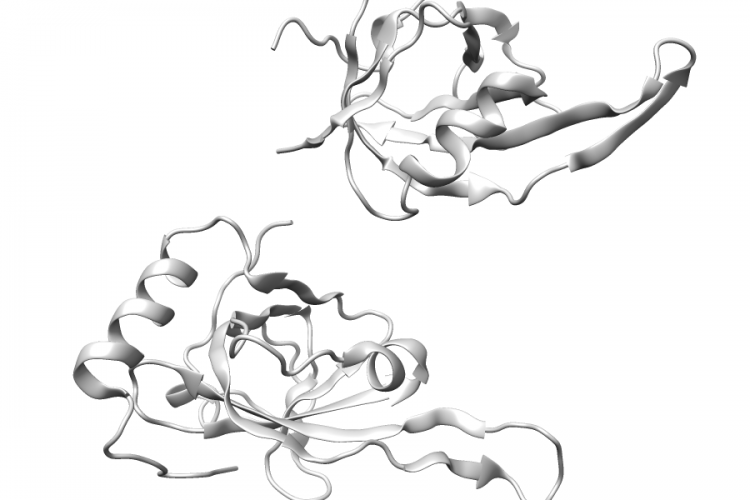
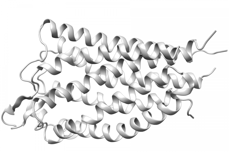
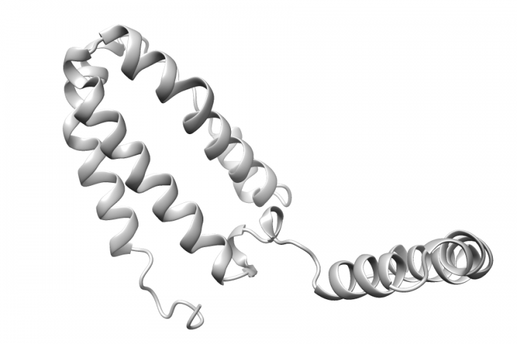
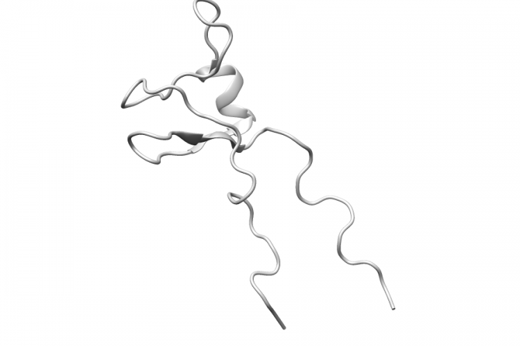
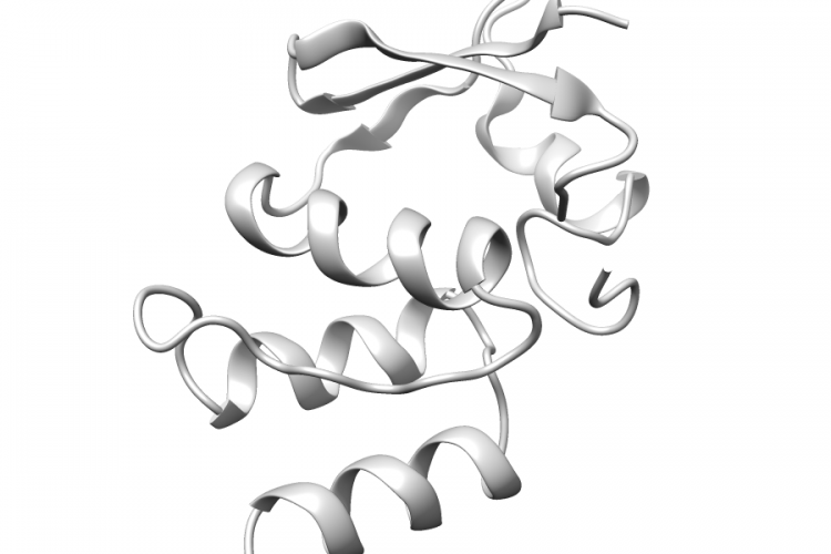
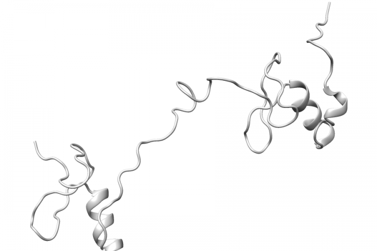
4BGC.A | KCNA3

General Structure Information 4bgc KCNA3 t1 domain of the renal potassium channel kv1.3 1.2Å AUTH W.KREMER,M.WEYAND,A.WINKLMEIER,C.SCHREIER,H.R.KALBITZERTITL 1.2 A X-RAY STRUCTURE OF THE RENAL POTASSIUM CHANNEL KV1.3TITL 2 T1 DOMAIN.REF PROTEIN J. V. 32 533 2013REFN ISSN 1572-3887PMID 24114469DOI 10.1007/S10930-013-9513-2 Variant Set Distributions Ripley’s K Analysis Plots Variant Set Comparisons Pathogenic Proximity Analysis Mapped Variants
See Details
4HC7.A | GATA3

General Structure Information 4hc7 GATA3 crystal structure of the full dna binding domain of gata3-complex 2 2.65Å AUTH Y.CHEN,D.L.BATES,R.DEY,P.H.CHEN,A.C.MACHADO,AUTH 2 I.A.LAIRD-OFFRINGA,R.ROHS,L.CHENTITL DNA BINDING BY GATA TRANSCRIPTION FACTOR SUGGESTS MECHANISMSTITL 2 OF DNA LOOPING AND LONG-RANGE GENE REGULATION.REF CELL REP V. 2 1197 2012REFN ESSN 2211-1247PMID 23142663DOI 10.1016/J.CELREP.2012.10.012 Variant Set Distributions Ripley’s K Analysis Plots Variant […]

See Details
1FGU.B | RPA1

General Structure Information 1fgu RPA1 ssdna-binding domain of the large subunit of replication protein a 2.5Å AUTH E.BOCHKAREVA,V.BELEGU,S.KOROLEV,A.BOCHKAREVTITL STRUCTURE OF THE MAJOR SINGLE-STRANDED DNA-BINDINGTITL 2 DOMAIN OF REPLICATION PROTEIN A SUGGESTS A DYNAMICTITL 3 MECHANISM FOR DNA BINDING.REF EMBO J. V. 20 612 2001REFN ISSN 0261-4189PMID 11157767DOI 10.1093/EMBOJ/20.3.612 Variant Set Distributions Ripley’s K Analysis Plots […]

See Details
3VW7.A | F2R

General Structure Information 3vw7 F2R crystal structure of human protease-activated receptor 1 (par1) bound with antagonist vorapaxar at 2.2 angstrom 2.2Å AUTH C.ZHANG,Y.SRINIVASAN,D.H.ARLOW,J.J.FUNG,D.PALMER,Y.ZHENG,AUTH 2 H.F.GREEN,A.PANDEY,R.O.DROR,D.E.SHAW,W.I.WEIS,S.R.COUGHLIN,AUTH 3 B.K.KOBILKATITL HIGH-RESOLUTION CRYSTAL STRUCTURE OF HUMANTITL 2 PROTEASE-ACTIVATED RECEPTOR 1REF NATURE V. 492 387 2012REFN ISSN 0028-0836PMID 23222541DOI 10.1038/NATURE11701 Variant Set Distributions Mapped Variants

See Details
1Y6K.L | IL10

General Structure Information 1y6k IL10 crystal structure of human il-10 complexed with the soluble il-10r1 chain 2.52Å AUTH S.I.YOON,B.C.JONES,N.J.LOGSDON,M.R.WALTERTITL SAME STRUCTURE, DIFFERENT FUNCTION CRYSTALTITL 2 STRUCTURE OF THE EPSTEIN-BARR VIRUS IL-10 BOUND TOTITL 3 THE SOLUBLE IL-10R1 CHAIN.REF STRUCTURE V. 13 551 2005REFN ISSN 0969-2126PMID 15837194DOI 10.1016/J.STR.2005.01.016 Variant Set Distributions Mapped Variants

See Details
1VD4.A | GTF2E1

General Structure Information 1vd4 GTF2E1 solution structure of the zinc finger domain of tfiie alpha -1.0Å AUTH M.OKUDA,A.TANAKA,Y.ARAI,M.SATOH,H.OKAMURA,AUTH 2 A.NAGADOI,F.HANAOKA,Y.OHKUMA,Y.NISHIMURATITL A NOVEL ZINC FINGER STRUCTURE IN THE LARGE SUBUNITTITL 2 OF HUMAN GENERAL TRANSCRIPTION FACTOR TFIIE.REF J.BIOL.CHEM. V. 279 51395 2004REFN ISSN 0021-9258PMID 15385556DOI 10.1074/JBC.M404722200 Variant Set Distributions Mapped Variants

See Details
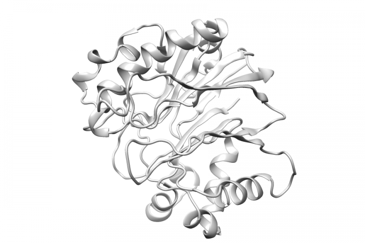
4AWN.A | DNASE1

General Structure Information 4awn DNASE1 structure of recombinant human dnase i (rhdnasei) in complex with magnesium and phosphate. 1.95Å AUTH G.PARSIEGLA,C.NOGUERE,L.SANTELL,R.A.LAZARUS,Y.BOURNETITL THE STRUCTURE OF HUMAN DNASE I BOUND TO MAGNESIUM ANDTITL 2 PHOSPHATE IONS POINTS TO A CATALYTIC MECHANISM COMMON TOTITL 3 MEMBERS OF THE DNASE I-LIKE SUPERFAMILY.REF BIOCHEMISTRY V. 51 10250 2012REFN ISSN 0006-2960PMID […]

See Details
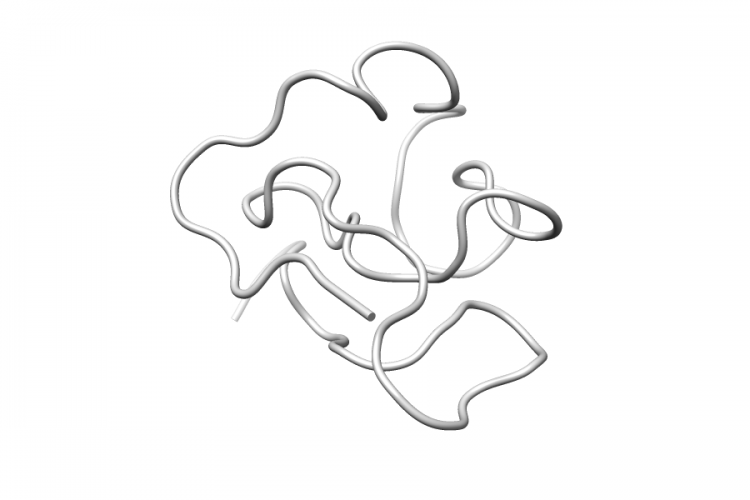
1BOR.A | PML

General Structure Information 1bor PML transcription factor pml, a proto-oncoprotein, nmr, 1 representative structure at ph 7.5, 30 c, in the presence of zinc -1.0Å AUTH K.L.BORDEN,M.N.BODDY,J.LALLY,N.J.OREILLY,S.MARTIN,AUTH 2 K.HOWE,E.SOLOMON,P.S.FREEMONTTITL THE SOLUTION STRUCTURE OF THE RING FINGER DOMAINTITL 2 FROM THE ACUTE PROMYELOCYTIC LEUKAEMIATITL 3 PROTO-ONCOPROTEIN PML.REF EMBO J. V. 14 1532 1995REFN ISSN 0261-4189PMID 7729428 […]

See Details
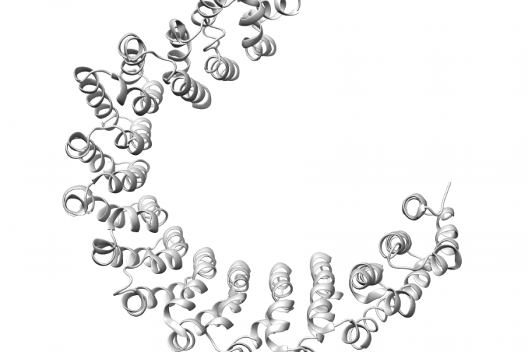
2NPP.A | PPP2R1A

General Structure Information 2npp PPP2R1A structure of the protein phosphatase 2a holoenzyme 3.3Å AUTH Y.XU,Y.XING,Y.CHEN,Y.CHAO,Z.LIN,E.FAN,J.W.YU,S.STRACK,AUTH 2 P.D.JEFFREY,Y.SHITITL STRUCTURE OF THE PROTEIN PHOSPHATASE 2A HOLOENZYMEREF CELL(CAMBRIDGE,MASS.) V. 127 1239 2006REFN ISSN 0092-8674PMID 17174897DOI 10.1016/J.CELL.2006.11.033 Variant Set Distributions Ripley’s K Analysis Plots Variant Set Comparisons Mapped Variants

See Details
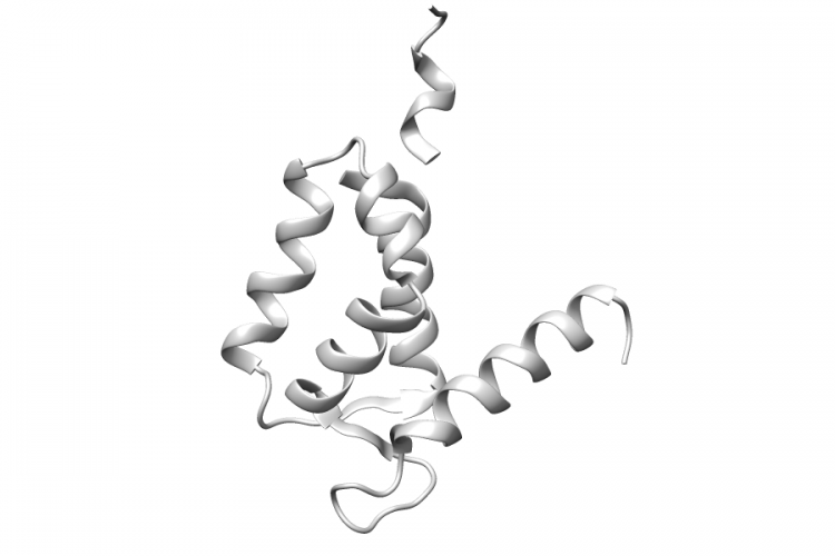
2LLT.A | S100A1

General Structure Information 2llt S100A1 post-translational s-nitrosylation is an endogenous factor fine-tuning human s100a1 protein properties -1.0Å AUTH M.LENARCIC ZIVKOVIC,M.ZAREBA-KOZIOL,L.ZHUKOVA,J.POZNANSKI,AUTH 2 I.ZHUKOV,A.WYSLOUCH-CIESZYNSKATITL POST-TRANSLATIONAL S-NITROSYLATION IS AN ENDOGENOUS FACTORTITL 2 FINE TUNING THE PROPERTIES OF HUMAN S100A1 PROTEIN.REF J.BIOL.CHEM. V. 287 40457 2012REFN ISSN 0021-9258PMID 22989881DOI 10.1074/JBC.M112.418392 Variant Set Distributions Mapped Variants

See Details