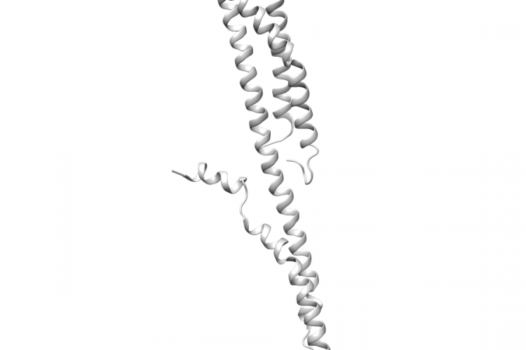
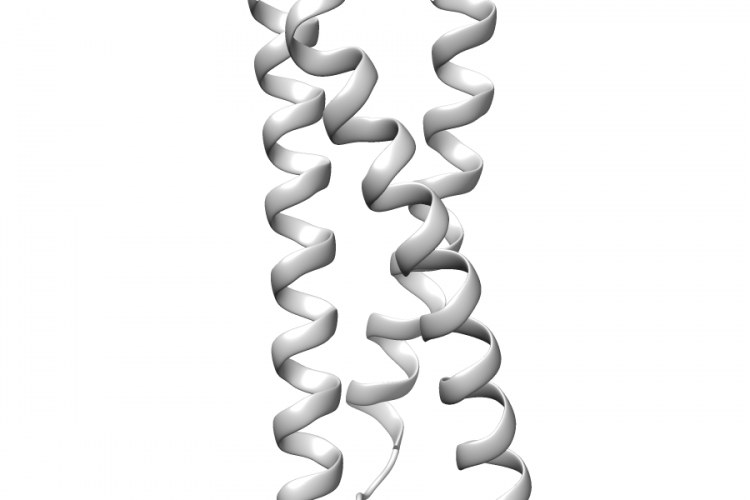
3LBX.B | SPTB

General Structure Information 3lbx SPTB crystal structure of the erythrocyte spectrin tetramerization domain complex 2.8Å AUTH J.J.IPSARO,S.L.HARPER,T.E.MESSICK,R.MARMORSTEIN,A.MONDRAGON,AUTH 2 D.W.SPEICHERTITL CRYSTAL STRUCTURE AND FUNCTIONAL INTERPRETATION OF THETITL 2 ERYTHROCYTE SPECTRIN TETRAMERIZATION DOMAIN COMPLEX.REF BLOOD V. 115 4843 2010REFN ISSN 0006-4971PMID 20197550DOI 10.1182/BLOOD-2010-01-261396 Variant Set Distributions Pathogenic Proximity Analysis Mapped Variants
See Details
1WRD.A | TOM1

General Structure Information 1wrd TOM1 crystal structure of tom1 gat domain in complex with ubiquitin 1.75Å AUTH M.AKUTSU,M.KAWASAKI,Y.KATOH,T.SHIBA,Y.YAMAGUCHI,R.KATO,AUTH 2 K.KATO,K.NAKAYAMA,S.WAKATSUKITITL STRUCTURAL BASIS FOR RECOGNITION OF UBIQUITINATED CARGO BYTITL 2 TOM1-GAT DOMAIN.REF FEBS LETT. V. 579 5385 2005REFN ISSN 0014-5793PMID 16199040DOI 10.1016/J.FEBSLET.2005.08.076 Variant Set Distributions Mapped Variants

See Details
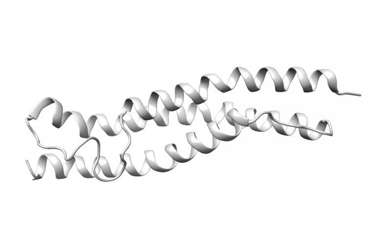
1WLX.A | ACTN4

General Structure Information 1wlx ACTN4 solution structure of the third spectrin repeat of alpha- actinin-4 -1.0Å AUTH K.KOWALSKI,A.L.MERKEL,G.W.BOOKERTITL SOLUTION STRUCTURE OF THE THIRD SPECTRIN REPEAT OFTITL 2 ALPHA-ACTININ-4REF TO BE PUBLISHEDREFN Variant Set Distributions Ripley’s K Analysis Plots Variant Set Comparisons Pathogenic Proximity Analysis Mapped Variants
See Details