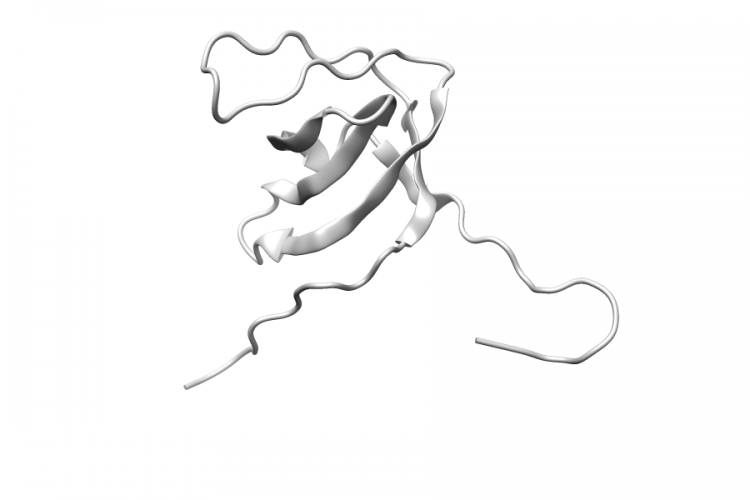
1X5O.A | RBMS1

General Structure Information 1x5o RBMS1 solution structure of rrm domain in rna binding motif, single-stranded interacting protein 1 -1.0Å AUTH W.DANG,Y.MUTO,M.INOUE,T.KIGAWA,M.SHIROUZU,T.TERADA,AUTH 2 S.YOKOYAMATITL SOLUTION STRUCTURE OF RRM DOMAIN IN RNA BINDINGTITL 2 MOTIF, SINGLE-STRANDED INTERACTING PROTEIN 1REF TO BE PUBLISHEDREFN Variant Set Distributions Mapped Variants
See Details
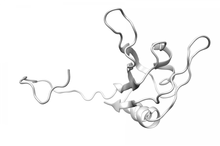
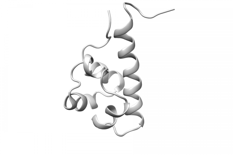
2E8N.A | EPHA2

General Structure Information 2e8n EPHA2 solution structure of the c-terminal sam-domain of ephaa2: ephrin type-a receptor 2 precursor (ec 2.7.10.1) -1.0Å AUTH A.K.GORONCY,M.SATO,S.KOSHIBA,M.INOUE,T.KIGAWA,AUTH 2 S.YOKOYAMATITL SOLUTION STRUCTURE OF THE C-TERMINAL SAM-DOMAIN OFTITL 2 EPHAA2: EPHRIN TYPE-A RECEPTOR 2 PRECURSOR (ECTITL 3 2.7.10.1)REF TO BE PUBLISHEDREFN Variant Set Distributions Mapped Variants
See Details
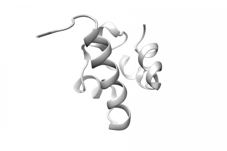
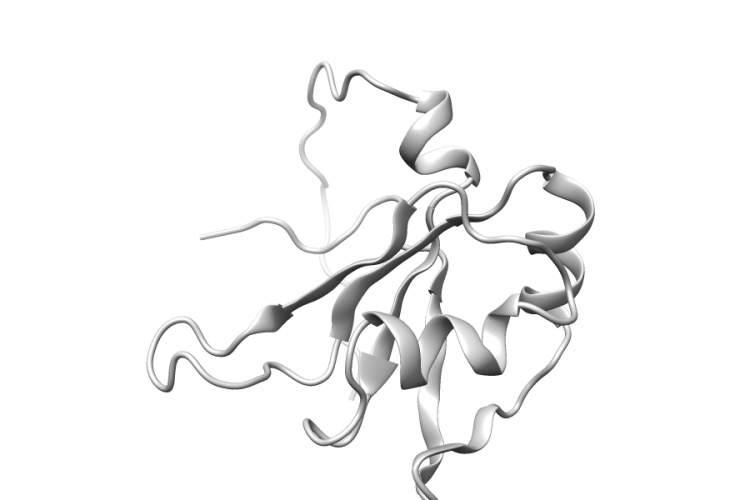
1UCV.A | EPHA8

General Structure Information 1ucv EPHA8 sterile alpha motif (sam) domain of ephrin type-a receptor 8 -1.0Å AUTH A.GORONCY,T.KIGAWA,S.KOSHIBA,N.KOBAYASHI,N.TOCHIO,AUTH 2 M.INOUE,S.YOKOYAMATITL NMR STRUCTURE OF STERILE ALPHA MOTIF (SAM) DOMAINTITL 2 OF EPHRIN TYPE-A RECEPTOR 8REF TO BE PUBLISHEDREFN Variant Set Distributions Mapped Variants
See Details
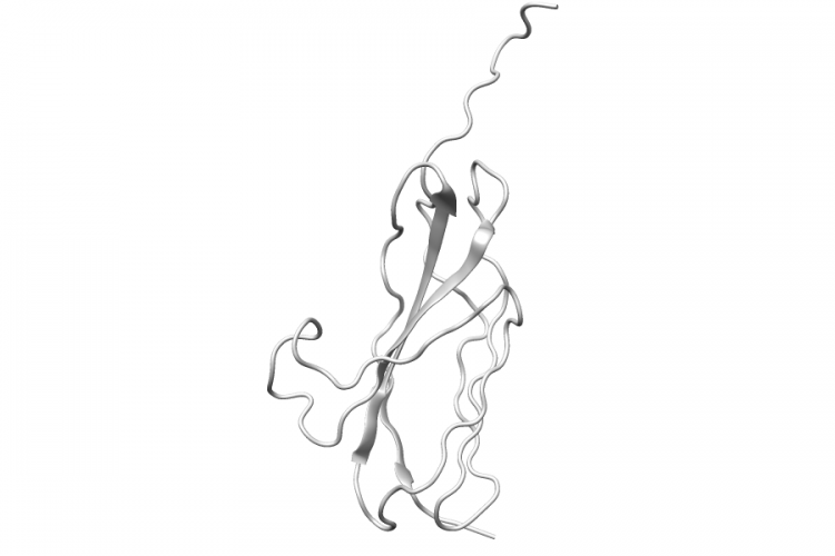
1X4B.A | HNRNPA2B1

General Structure Information 1x4b HNRNPA2B1 solution structure of rrm domain in heterogeneous nuclear ribonucleaoproteins a2/b1 -1.0Å AUTH F.HE,Y.MUTO,M.INOUE,T.KIGAWA,M.SHIROUZU,T.TERADA,AUTH 2 S.YOKOYAMATITL SOLUTION STRUCTURE OF RRM DOMAIN IN HETEROGENEOUSTITL 2 NUCLEAR RIBONUCLEAOPROTEINS A2/B1REF TO BE PUBLISHEDREFN Variant Set Distributions Mapped Variants
See Details
2DLH.A | PTPRD

General Structure Information 2dlh PTPRD solution structure of the second fn3 domain of human receptor-type tyrosine-protein phosphatase delta -1.0Å AUTH K.SEIMIYA,F.HAYASHI,M.YOSHIDA,S.YOKOYAMATITL SOLUTION STRUCTURE OF THE SECOND FN3 DOMAIN OFTITL 2 HUMAN RECEPTOR-TYPE TYROSINE-PROTEIN PHOSPHATASETITL 3 DELTAREF TO BE PUBLISHEDREFN Variant Set Distributions Ripley’s K Analysis Plots Variant Set Comparisons Pathogenic Proximity Analysis Mapped Variants

See Details
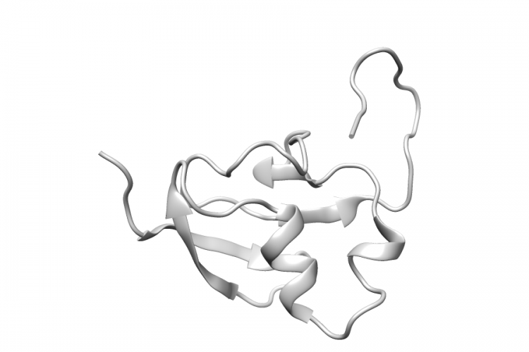
1WWT.A | TARS

General Structure Information 1wwt TARS solution structure of the tgs domain from human threonyl- trna synthetase -1.0Å AUTH E.CHIKAYAMA,T.KIGAWA,T.TOMIZAWA,S.KOSHIBA,M.INOUE,AUTH 2 S.YOKOYAMATITL SOLUTION STRUCTURE OF THE TGS DOMAIN FROM HUMANTITL 2 THREONYL-TRNA SYNTHETASEREF TO BE PUBLISHEDREFN Variant Set Distributions Mapped Variants
See Details
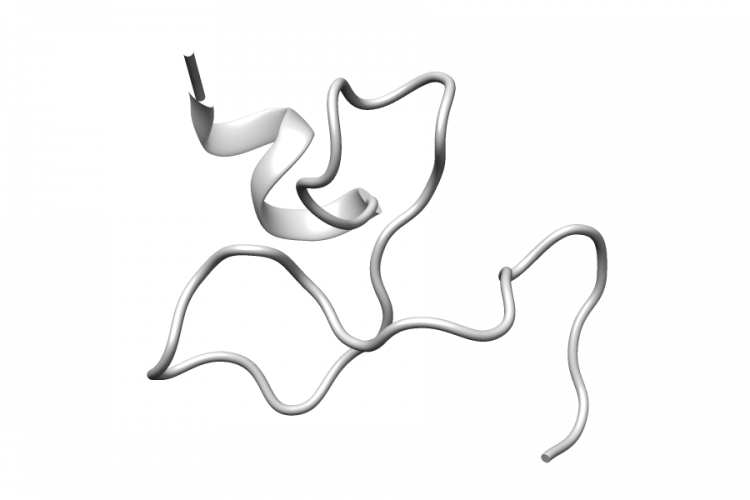
2EQE.A | TNFAIP3

General Structure Information 2eqe TNFAIP3 solution structure of the fourth a20-type zinc finger domain from human tumor necrosis factor, alpha-induced protein3 -1.0Å AUTH H.P.ZHANG,T.NAGASIMA,F.HAYASHI,S.YOKOYAMATITL SOLUTION STRUCTURE OF THE FOURTH A20-TYPE ZINCTITL 2 FINGER DOMAIN FROM HUMAN TUMOR NECROSIS FACTOR,TITL 3 ALPHA-INDUCED PROTEIN3REF TO BE PUBLISHEDREFN Variant Set Distributions Mapped Variants
See Details
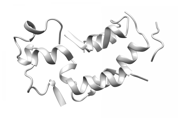
1X0J.A | BRD2

General Structure Information 1x0j BRD2 crystal structure analysis of the n-terminal bromodomain of human brd2 1.8Å AUTH Y.NAKAMURA,T.UMEHARA,M.SHIROUZU,B.PADMANABHAN,AUTH 2 S.YOKOYAMATITL STRUCTURAL BASIS FOR ACETYLATED HISTONE H4TITL 2 RECOGNITION BY THE HUMAN BRD2 BROMODOMAINREF TO BE PUBLISHEDREFN Variant Set Distributions Mapped Variants
See Details
2D8M.A | XRCC1

General Structure Information 2d8m XRCC1 solution structure of the first brct domain of dna-repair protein xrcc1 -1.0Å AUTH T.NAGASHIMA,F.HAYASHI,S.YOKOYAMATITL SOLUTION STRUCTURE OF THE FIRST BRCT DOMAIN OFTITL 2 DNA-REPAIR PROTEIN XRCC1REF TO BE PUBLISHEDREFN Variant Set Distributions Ripley’s K Analysis Plots Variant Set Comparisons Pathogenic Proximity Analysis Mapped Variants

See Details
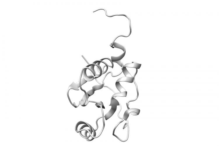
2CUB.A | NCK1

General Structure Information 2cub NCK1 solution structure of the sh3 domain of the human cytoplasmic protein nck1 -1.0Å AUTH S.OHNISHI,T.KIGAWA,M.SATO,T.TOMIZAWA,S.KOSHIBA,AUTH 2 M.INOUE,S.YOKOYAMATITL SOLUTION STRUCTURE OF THE SH3 DOMAIN OF THE HUMANTITL 2 CYTOPLASMIC PROTEIN NCK1REF TO BE PUBLISHEDREFN Variant Set Distributions Mapped Variants
See Details