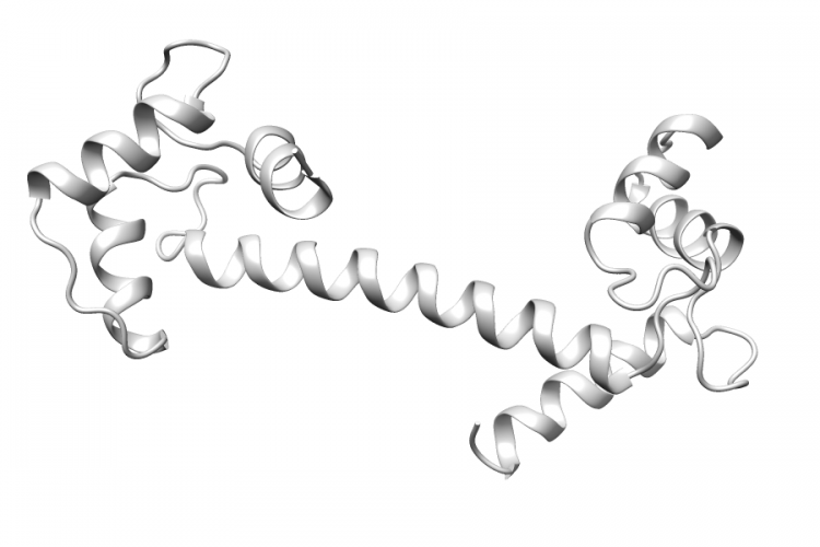
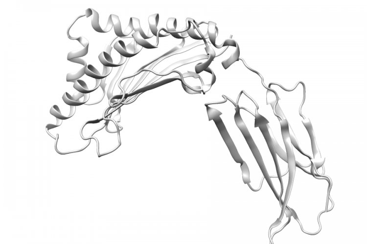
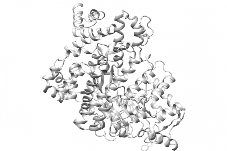
3CJW.A | NR2F2

General Structure Information 3cjw NR2F2 crystal structure of the human coup-tfii ligand binding domain 1.48Å AUTH S.W.KRUSE,K.SUINO-POWELL,X.E.ZHOU,J.E.KRETSCHMAN,AUTH 2 R.REYNOLDS,C.VONRHEIN,Y.XU,L.WANG,S.Y.TSAI,AUTH 3 M.J.TSAI,H.E.XUTITL IDENTIFICATION OF COUP-TFII ORPHAN NUCLEARTITL 2 RECEPTOR AS A RETINOIC ACID-ACTIVATED RECEPTOR.REF PLOS BIOL. V. 6 E227 2008REFN ISSN 1544-9173PMID 18798693DOI 10.1371/JOURNAL.PBIO.0060227 Variant Set Distributions Ripley’s K Analysis Plots Variant Set Comparisons Pathogenic Proximity […]
See Details
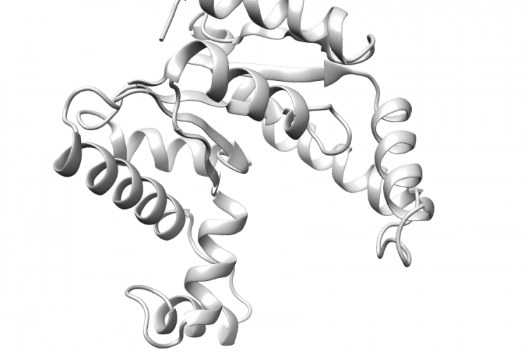
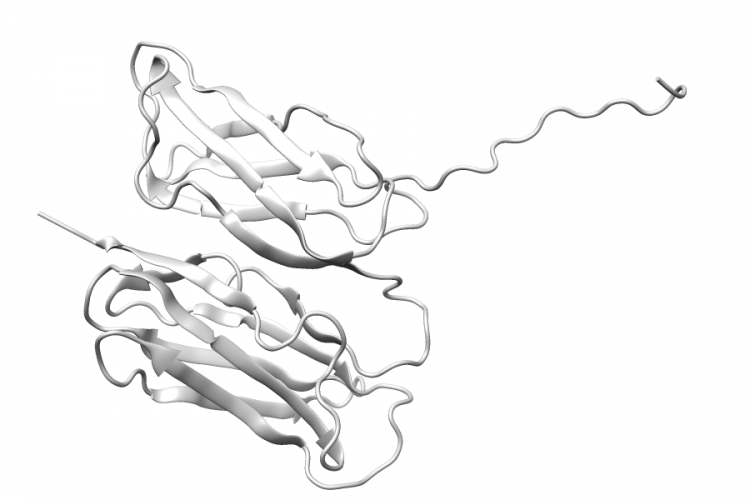
3HNC.A | RRM1

General Structure Information 3hnc RRM1 crystal structure of human ribonucleotide reductase 1 bound to the effector ttp 2.41Å AUTH J.W.FAIRMAN,S.R.WIJERATHNA,M.F.AHMAD,H.XU,R.NAKANO,S.JHA,AUTH 2 J.PRENDERGAST,R.M.WELIN,S.FLODIN,A.ROOS,P.NORDLUND,Z.LI,AUTH 3 T.WALZ,C.G.DEALWISTITL STRUCTURAL BASIS FOR ALLOSTERIC REGULATION OF HUMANTITL 2 RIBONUCLEOTIDE REDUCTASE BY NUCLEOTIDE-INDUCEDTITL 3 OLIGOMERIZATION.REF NAT.STRUCT.MOL.BIOL. V. 18 316 2011REFN ISSN 1545-9993PMID 21336276DOI 10.1038/NSMB.2007 Variant Set Distributions Ripley’s K Analysis Plots Variant […]

See Details
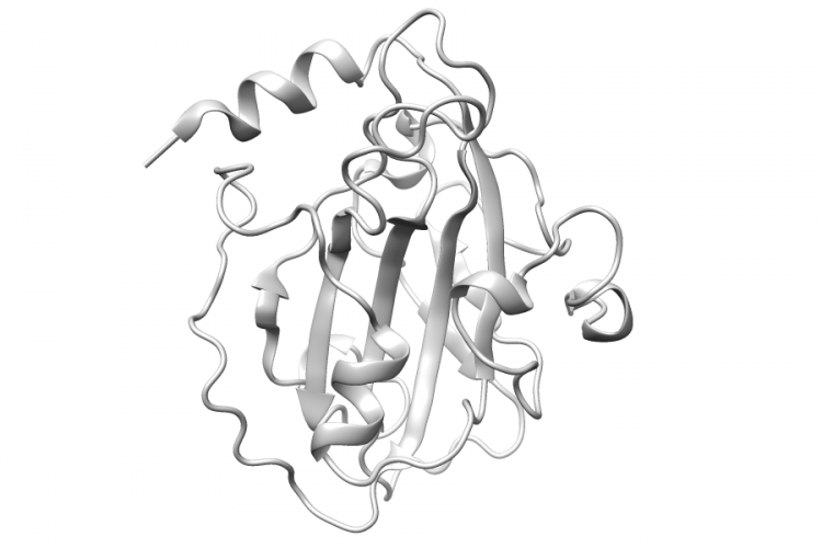
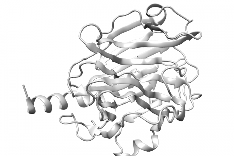
1GGZ.A | CALML3

General Structure Information 1ggz CALML3 crystal structure of the calmodulin-like protein (hclp) from human epithelial cells 1.5Å AUTH B.G.HAN,M.HAN,H.SUI,P.YASWEN,P.J.WALIAN,B.K.JAPTITL CRYSTAL STRUCTURE OF HUMAN CALMODULIN-LIKETITL 2 PROTEIN: INSIGHTS INTO ITS FUNCTIONAL ROLE.REF FEBS LETT. V. 521 24 2002REFN ISSN 0014-5793PMID 12067719DOI 10.1016/S0014-5793(02)02780-1 Variant Set Distributions Ripley’s K Analysis Plots Variant Set Comparisons Pathogenic Proximity Analysis Mapped […]

See Details
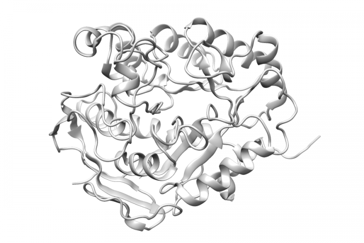
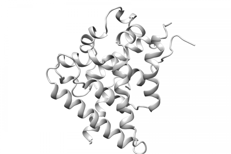
1TEV.A | CMPK1

General Structure Information 1tev CMPK1 crystal structure of the human ump/cmp kinase in open conformation 2.1Å AUTH N.SEKULIC,S.ORT,M.KONRAD,A.LAVIETITL SUBSTRATE-INDUCED CONFORMATIONAL CHANGES IN HUMANTITL 2 UMP/CMP KINASE.REF J.BIOL.CHEM. V. 279 33882 2004REFN ISSN 0021-9258PMID 15163660DOI 10.1074/JBC.M401989200 Variant Set Distributions Ripley’s K Analysis Plots Variant Set Comparisons Pathogenic Proximity Analysis Mapped Variants

See Details
1BD9.B | PEBP1

General Structure Information 1bd9 PEBP1 human phosphatidylethanolamine binding protein 2.05Å AUTH M.J.BANFIELD,J.J.BARKER,A.C.PERRY,R.L.BRADYTITL FUNCTION FROM STRUCTURE? THE CRYSTAL STRUCTURE OFTITL 2 HUMAN PHOSPHATIDYLETHANOLAMINE-BINDING PROTEINTITL 3 SUGGESTS A ROLE IN MEMBRANE SIGNAL TRANSDUCTION.REF STRUCTURE V. 6 1245 1998REFN ISSN 0969-2126PMID 9782050DOI 10.1016/S0969-2126(98)00125-7 Variant Set Distributions Mapped Variants

See Details
4C6F.A | CAD

General Structure Information 4c6f CAD crystal structure of the dihydroorotase domain of human cad bound to substrate at ph 6.5 1.26Å AUTH A.GRANDE-GARCIA,N.LALLOUS,C.DIAZ-TEJADA,S.RAMON-MAIQUESTITL STRUCTURE, FUNCTIONAL CHARACTERIZATION AND EVOLUTION OF THETITL 2 DIHYDROOROTASE DOMAIN OF HUMAN CAD.REF STRUCTURE V. 22 185 2014REFN ISSN 0969-2126PMID 24332717DOI 10.1016/J.STR.2013.10.016 Variant Set Distributions Ripley’s K Analysis Plots Variant Set Comparisons […]

See Details
1T7Y.A | AZGP1

General Structure Information 1t7y AZGP1 zn-alpha-2-glycoprotein; baculo-zag peg 200, no glycerol 2.8Å AUTH S.L.DELKER,A.P.WEST JR.,L.MCDERMOTT,M.W.KENNEDY,P.J.BJORKMANTITL CRYSTALLOGRAPHIC STUDIES OF LIGAND BINDING BYTITL 2 ZN-ALPHA2-GLYCOPROTEIN.REF J.STRUCT.BIOL. V. 148 205 2004REFN ISSN 1047-8477PMID 15477100DOI 10.1016/J.JSB.2004.04.009 Variant Set Distributions Ripley’s K Analysis Plots Variant Set Comparisons Pathogenic Proximity Analysis Mapped Variants

See Details
2K7P.A | FLNA

General Structure Information 2k7p FLNA filamin a ig-like domains 16-17 -1.0Å AUTH O.K.HEIKKINEN,S.RUSKAMO,P.V.KONAREV,D.I.SVERGUN,AUTH 2 T.IIVANAINEN,S.M.HEIKKINEN,P.PERMI,H.KOSKELA,AUTH 3 I.KILPELAINEN,J.YLANNETITL ATOMIC STRUCTURES OF TWO NOVEL IMMUNOGLOBULIN-LIKETITL 2 DOMAIN PAIRS IN THE ACTIN CROSS-LINKING PROTEINTITL 3 FILAMINREF J.BIOL.CHEM. V. 284 25450 2009REFN ISSN 0021-9258PMID 19622754DOI 10.1074/JBC.M109.019661 Variant Set Distributions Mapped Variants

See Details
3POS.B | CALR

General Structure Information 3pos CALR crystal structure of the globular domain of human calreticulin 1.65Å AUTH A.CHOUQUET,H.PAIDASSI,W.L.LING,P.FRACHET,G.HOUEN,G.J.ARLAUD,AUTH 2 C.GABORIAUDTITL X-RAY STRUCTURE OF THE HUMAN CALRETICULIN GLOBULAR DOMAINTITL 2 REVEALS A PEPTIDE-BINDING AREA AND SUGGESTS ATITL 3 MULTI-MOLECULAR MECHANISMREF PLOS ONE V. 6 E1788 2011REFN ESSN 1932-6203PMID 21423620DOI 10.1371/JOURNAL.PONE.0017886 Variant Set Distributions Mapped Variants

See Details