General Structure Information
| PDB ID | 1rj7 |
| HGNC Gene Label(s) | EDA |
| Structure Name | crystal structure of eda-a1 |
| Resolution | 2.3Å |
| Reference | AUTH S.G.HYMOWITZ,D.M.COMPAAN,M.YAN,H.J.WALLWEBER,V.M.DIXIT,AUTH 2 M.A.STAROVASNIK,A.M.DE VOSTITL THE CRYSTAL STRUCTURES OF EDA-A1 AND EDA-A2: SPLICE VARIANTSTITL 2 WITH DISTINCT RECEPTOR SPECIFICITY.REF STRUCTURE V. 11 1513 2003REFN ISSN 0969-2126PMID 14656435DOI 10.1016/J.STR.2003.11.009 |
Variant Set Distributions
ExAC Variants
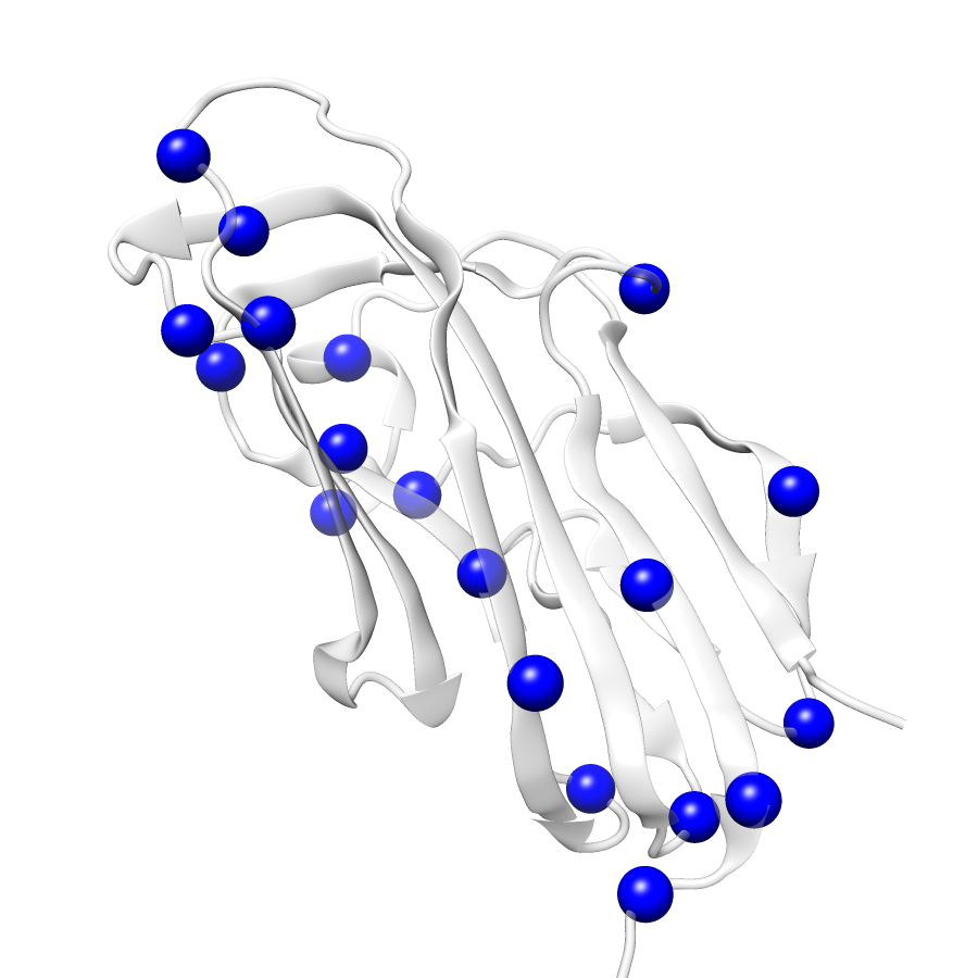
| Number Of Residues | 143 |
| Number Of SNVs | 19 |
| Number Of Permutations | 8282 |
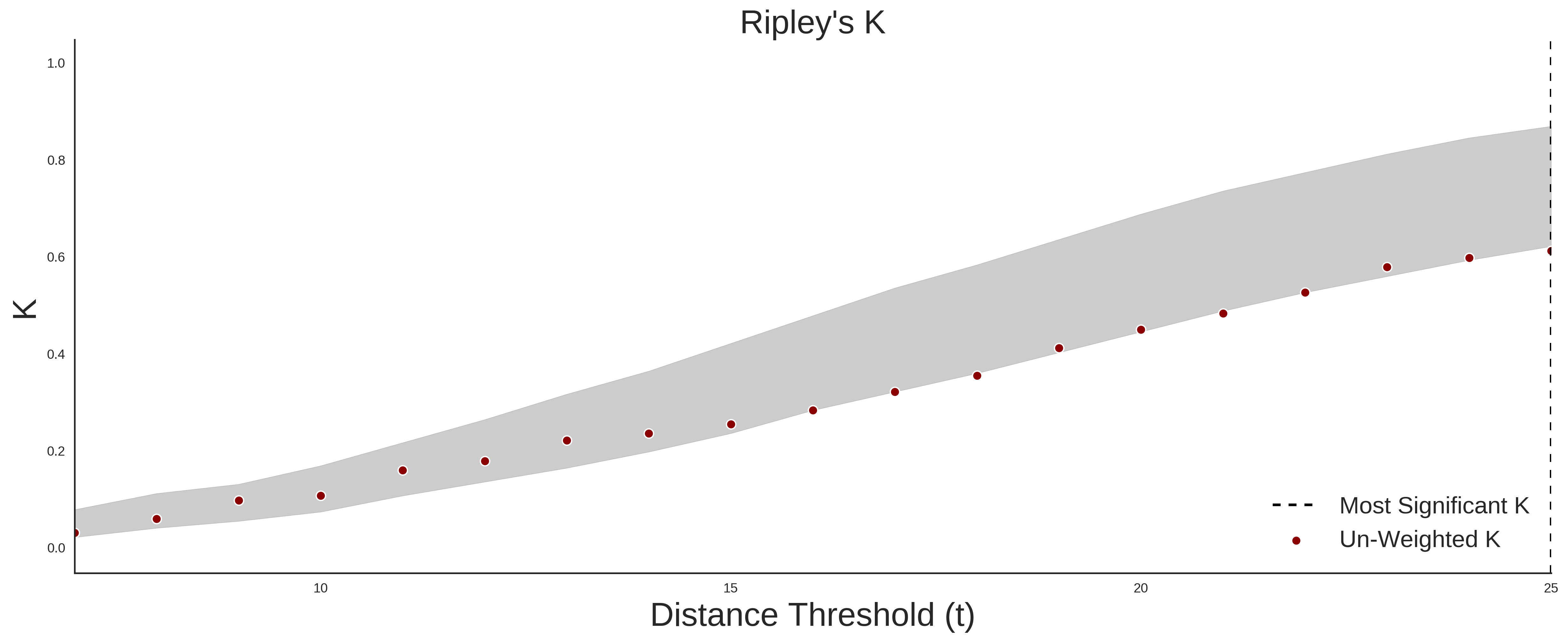
| Optimal Distance Threshold | 6.0 |
| K Statistic | 0.006 |
| p-value | 0.656 |
ClinVar
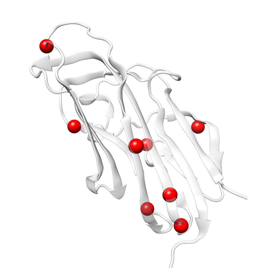
| Number Of Residues | 143 |
| Number Of SNVs | 8 |
| Number Of Permutations | 3336 |
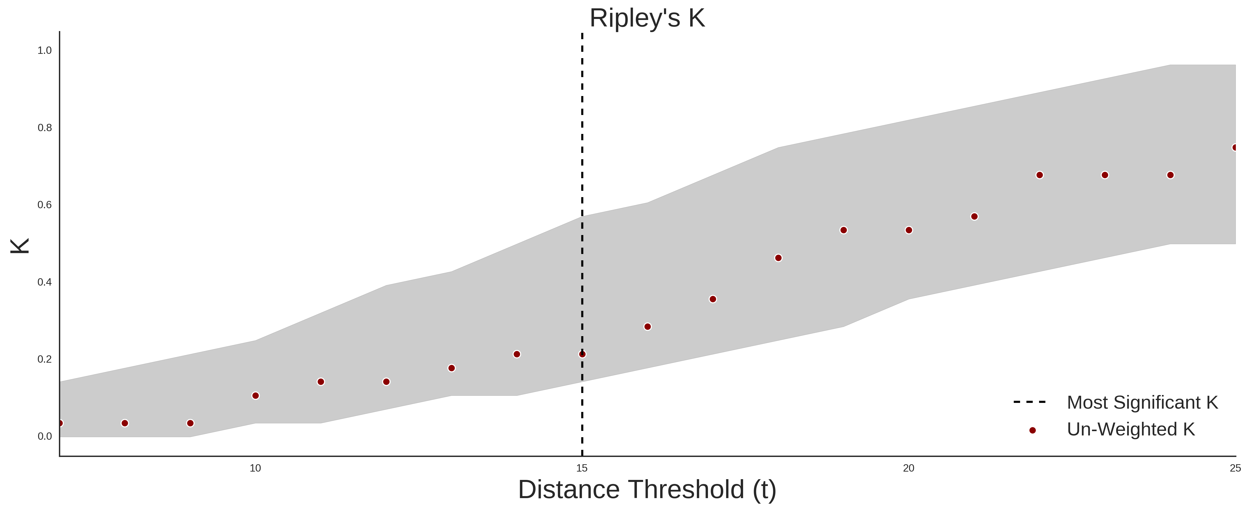
| Optimal Distance Threshold | 5.0 |
| K Statistic | 0.036 |
| p-value | 0.507 |
Ripley’s K Analysis Plots
ExACClinVar
Variant Set Comparisons
ClinVar vs. ExAC
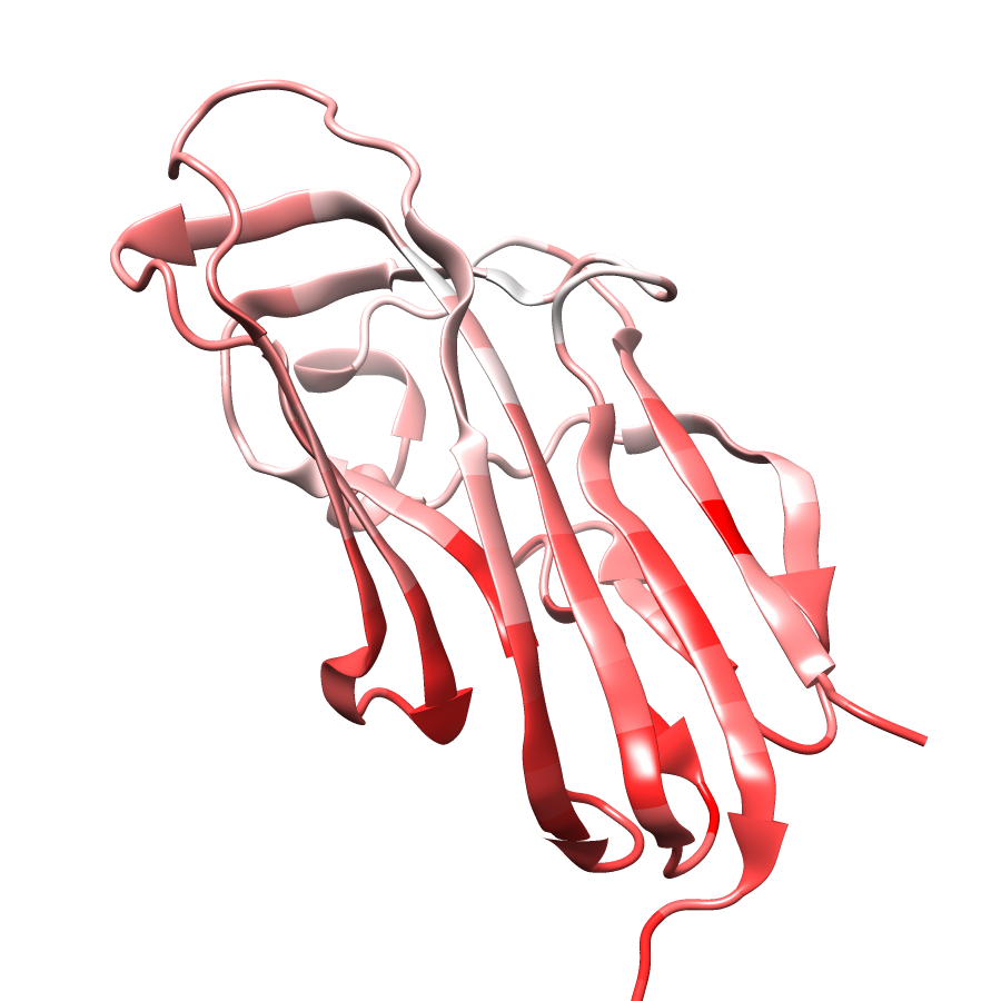
| Number Of ExAC SNVs | 19 |
| Number Of ClinVar SNVs | 8 |
| Optimal Distance Threshold | 5.0 |
| K Statistic | 0.036 |
| p-value | 0.743 |