General Structure Information
| PDB ID | 2wfj |
| HGNC Gene Label(s) | PPIG |
| Structure Name | atomic resolution crystal structure of the ppiase domain of human cyclophilin g in complex with cyclosporin a. |
| Resolution | 0.75Å |
| Reference | AUTH C.M.STEGMANN,D.SEELIGER,G.M.SHELDRICK,B.L.DE GROOT,M.C.WAHLTITL THE THERMODYNAMIC INFLUENCE OF TRAPPED WATER MOLECULES ON ATITL 2 PROTEIN-LIGAND INTERACTION.REF ANGEW.CHEM.INT.ED.ENGL. V. 48 5207 2009REFN ISSN 1433-7851PMID 19499554DOI 10.1002/ANIE.200900481 |
Variant Set Distributions
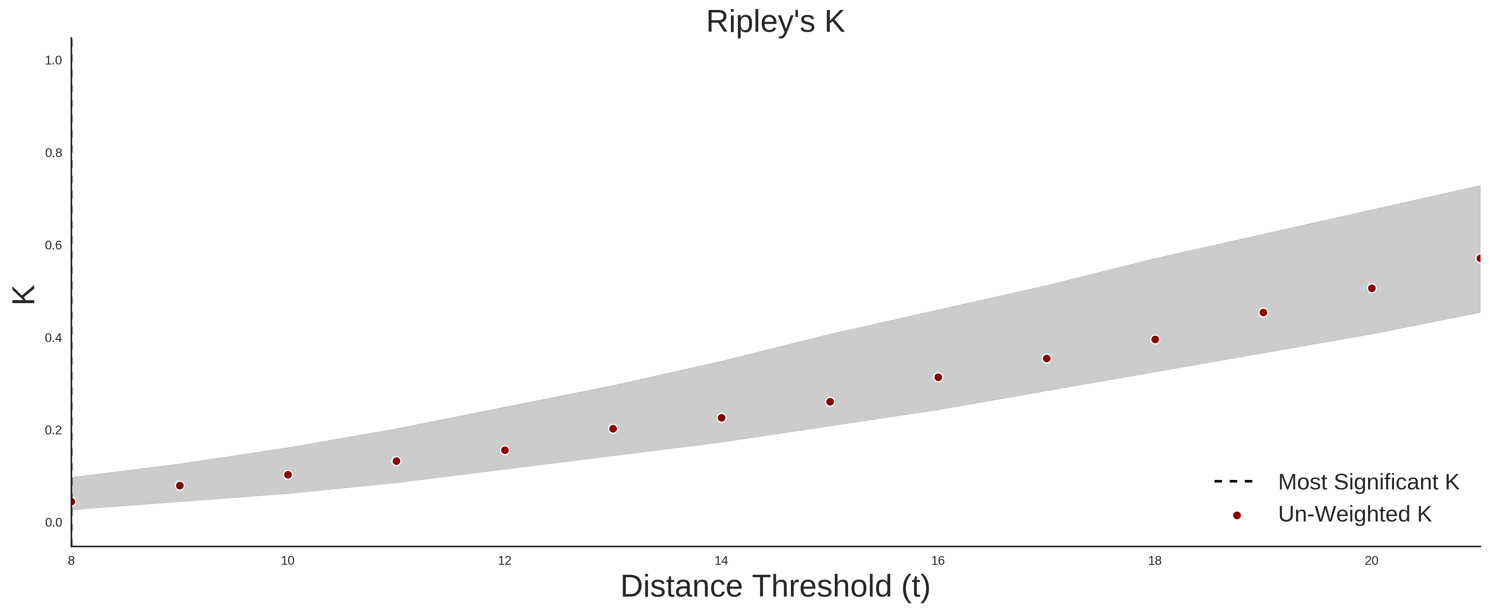
ExAC Variants
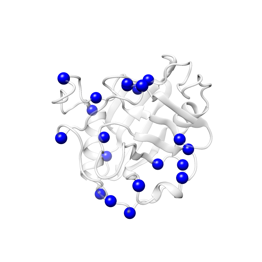
| Number Of Residues | 172 |
| Number Of SNVs | 19 |
| Number Of Permutations | 4494 |
| Optimal Distance Threshold | 8.0 |
| K Statistic | 0.047 |
| p-value | 0.84 |
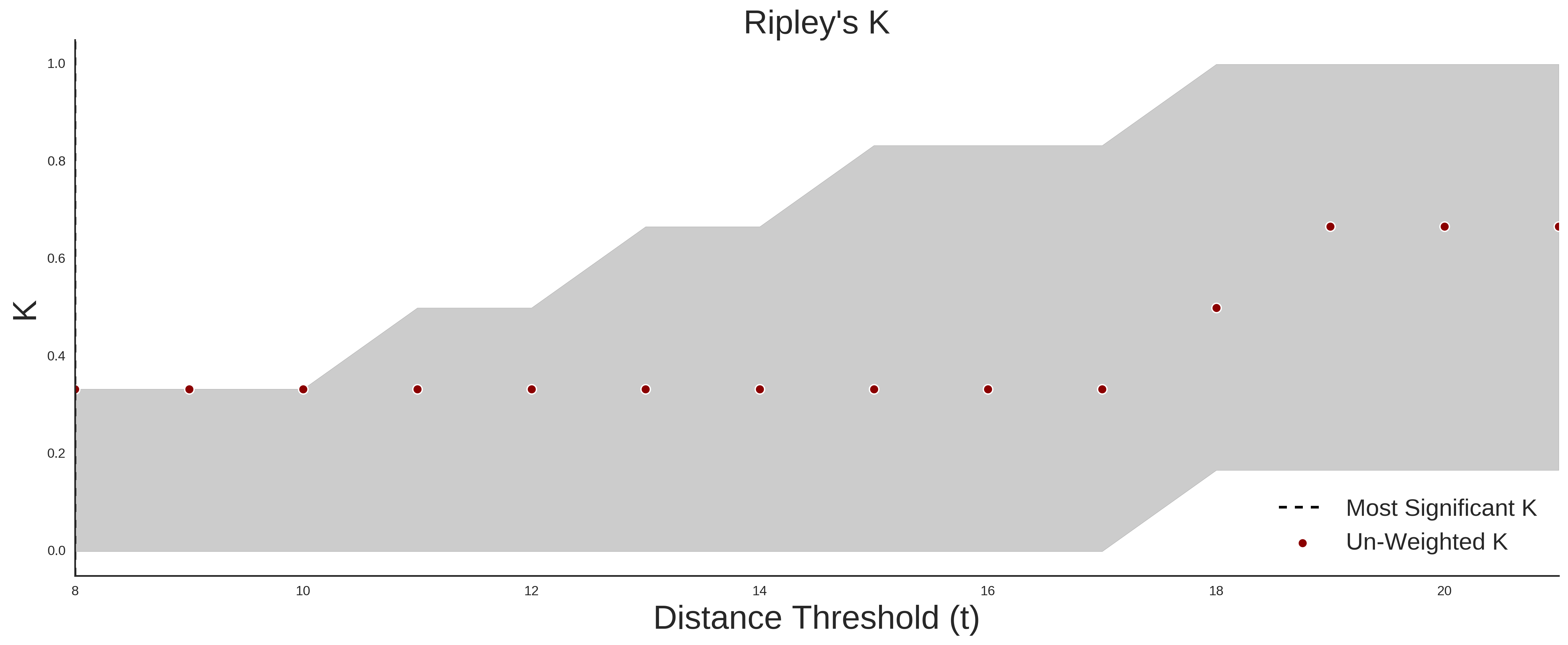
COSMIC
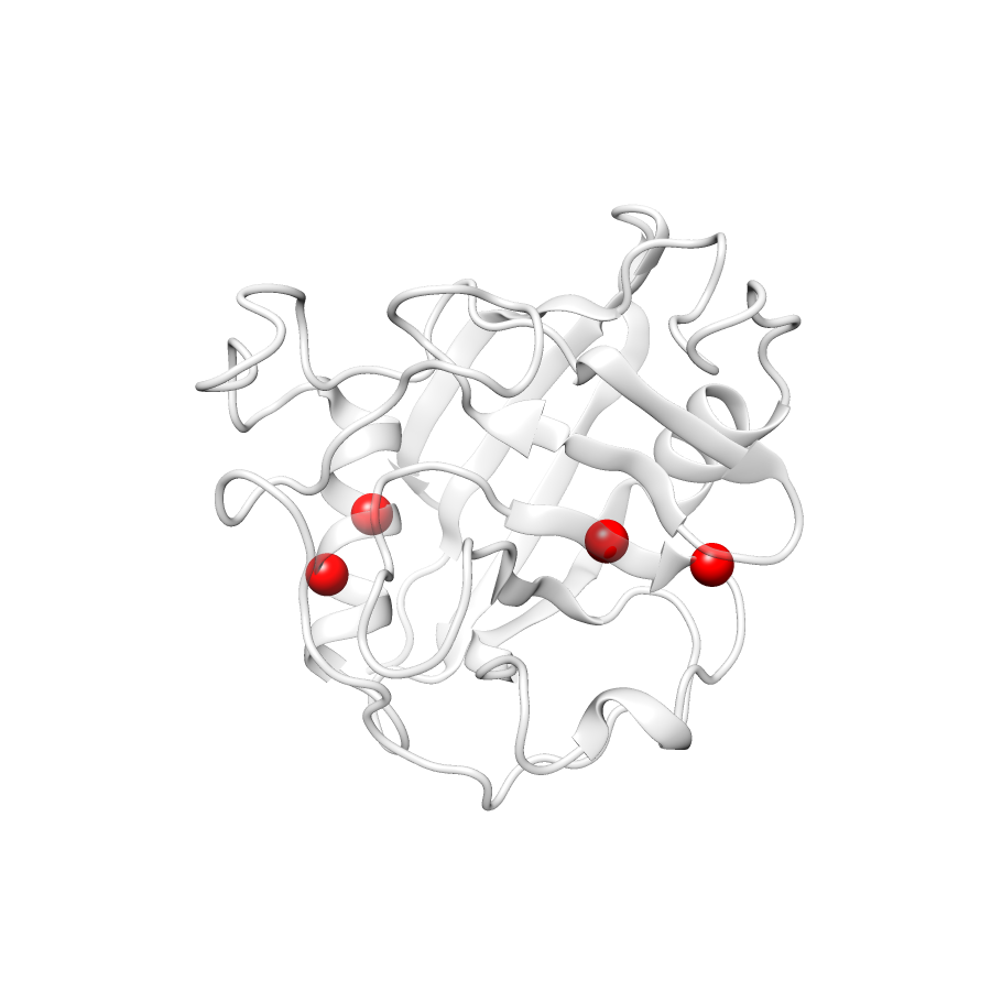
| Number Of Residues | 172 |
| Number Of SNVs | 4 |
| Number Of Permutations | 338 |
| Optimal Distance Threshold | 5.0 |
| K Statistic | 0.167 |
| p-value | 0.061 |
Ripley’s K Analysis Plots
ExACCOSMIC
Variant Set Comparisons
Cosmic vs. ExAC
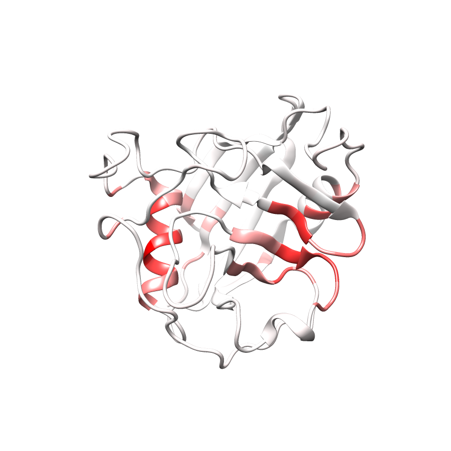
| Number Of ExAC SNVs | 19 |
| Number Of COSMIC SNVs | 4 |
| Optimal Distance Threshold | 5.0 |
| K Statistic | 0.161 |
| p-value | 0.263 |