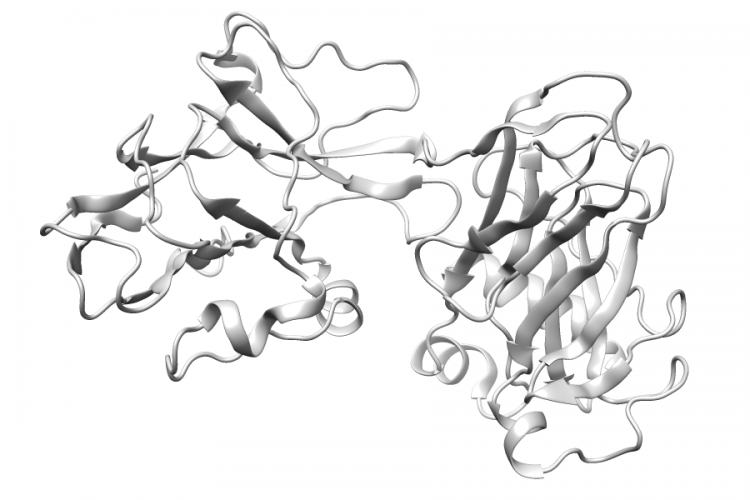
General Structure Information
| PDB ID | 3ud1 |
| HGNC Gene Label(s) | ANK1 |
| Structure Name | crystal structure of zu5a-zu5b domains of human erythrocyte ankyrin |
| Resolution | 2.0Å |
| Reference | AUTH M.YASUNAGA,J.J.IPSARO,A.MONDRAGONTITL STRUCTURALLY SIMILAR BUT FUNCTIONALLY DIVERSE ZU5 DOMAINS INTITL 2 HUMAN ERYTHROCYTE ANKYRIN.REF J.MOL.BIOL. V. 417 336 2012REFN ISSN 0022-2836PMID 22310050DOI 10.1016/J.JMB.2012.01.041 |
Variant Set Distributions
ExAC Variants
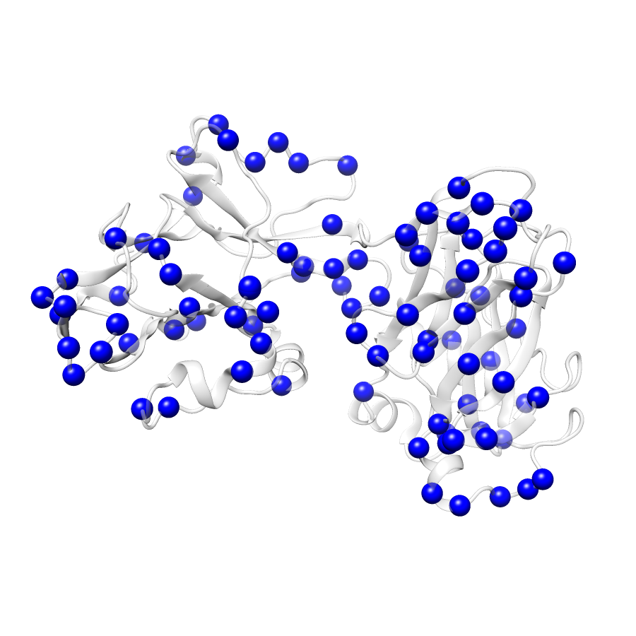
| Number Of Residues | 322 |
| Number Of SNVs | 87 |
| Number Of Permutations | 13740 |
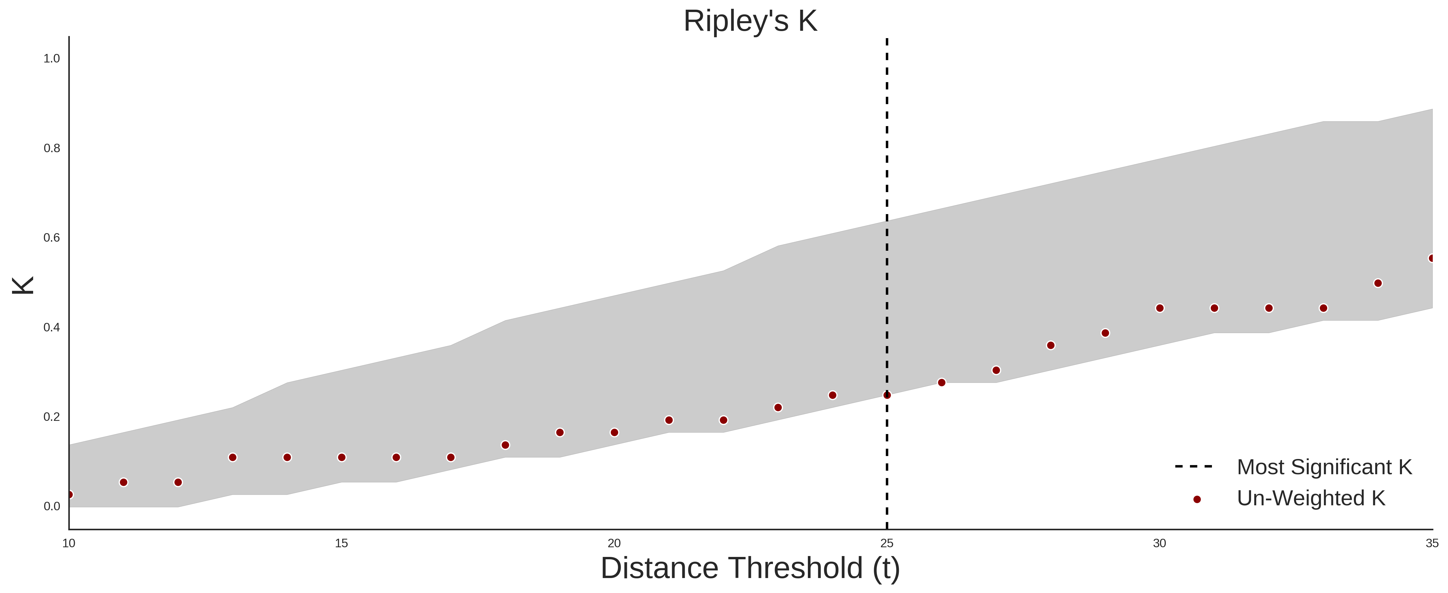
| Optimal Distance Threshold | 17.0 |
| K Statistic | 0.172 |
| p-value | 0.033 |
COSMIC
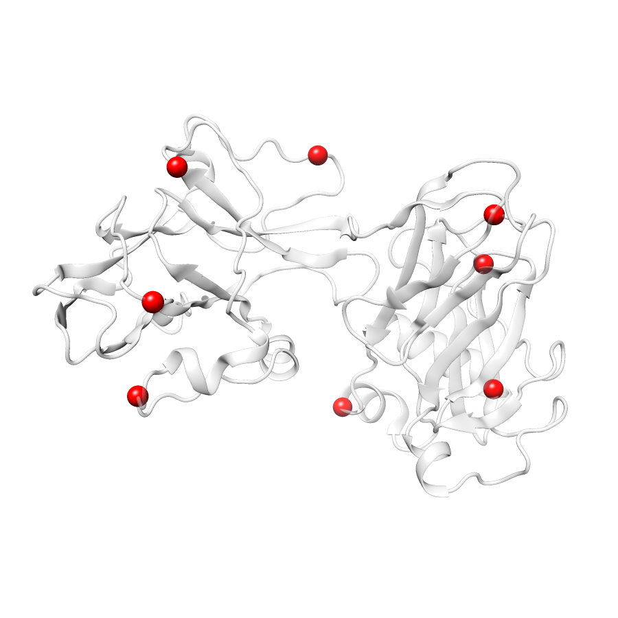
| Number Of Residues | 322 |
| Number Of SNVs | 8 |
| Number Of Permutations | 1807 |
| Optimal Distance Threshold | 17.0 |
| K Statistic | 0.071 |
| p-value | 0.085 |
Ripley’s K Analysis Plots
ExACCOSMIC
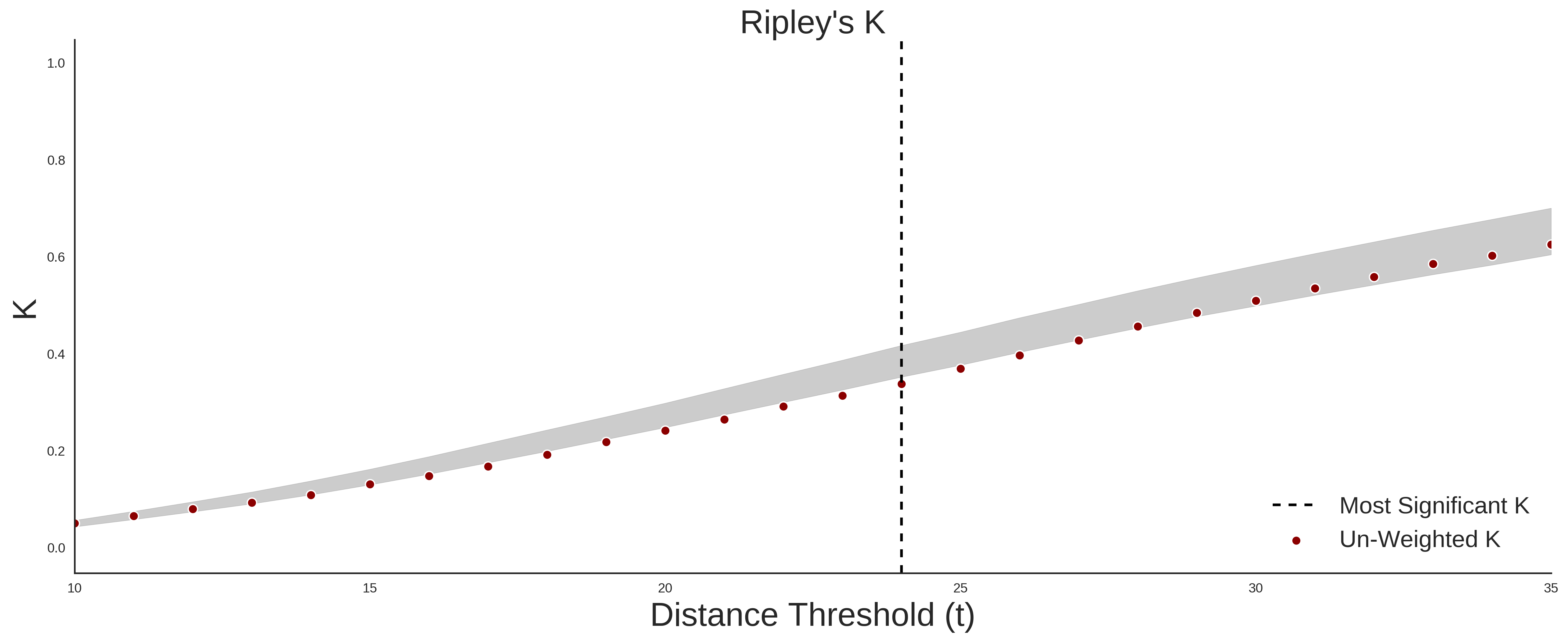
Variant Set Comparisons
Cosmic vs. ExAC
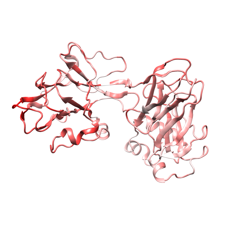
| Number Of ExAC SNVs | 87 |
| Number Of COSMIC SNVs | 8 |
| Optimal Distance Threshold | 17.0 |
| K Statistic | -0.1 |
| p-value | 0.164 |