General Structure Information
| PDB ID | 3kbt |
| HGNC Gene Label(s) | SPTB |
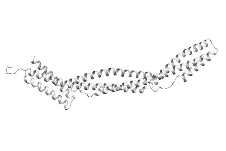
| Structure Name | crystal structure of the ankyrin binding domain of human erythroid beta spectrin (repeats 13-15) in complex with the spectrin binding domain of human erythroid ankyrin (zu5-ank) |
| Resolution | 2.75Å |
| Reference | AUTH J.J.IPSARO,A.MONDRAGONTITL STRUCTURAL BASIS FOR SPECTRIN RECOGNITION BY ANKYRIN.REF BLOOD V. 115 4093 2010REFN ISSN 0006-4971PMID 20101027DOI 10.1182/BLOOD-2009-11-255604 |
Variant Set Distributions
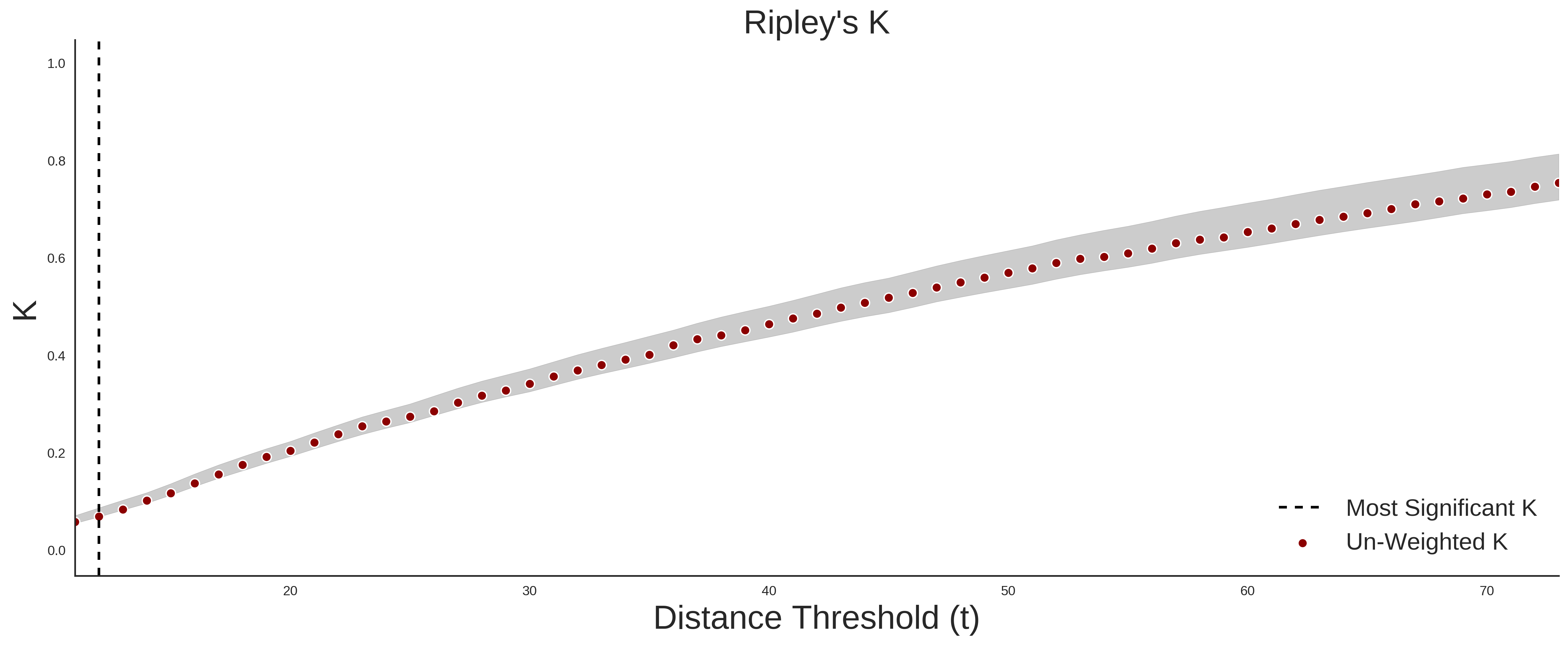
ExAC Variants
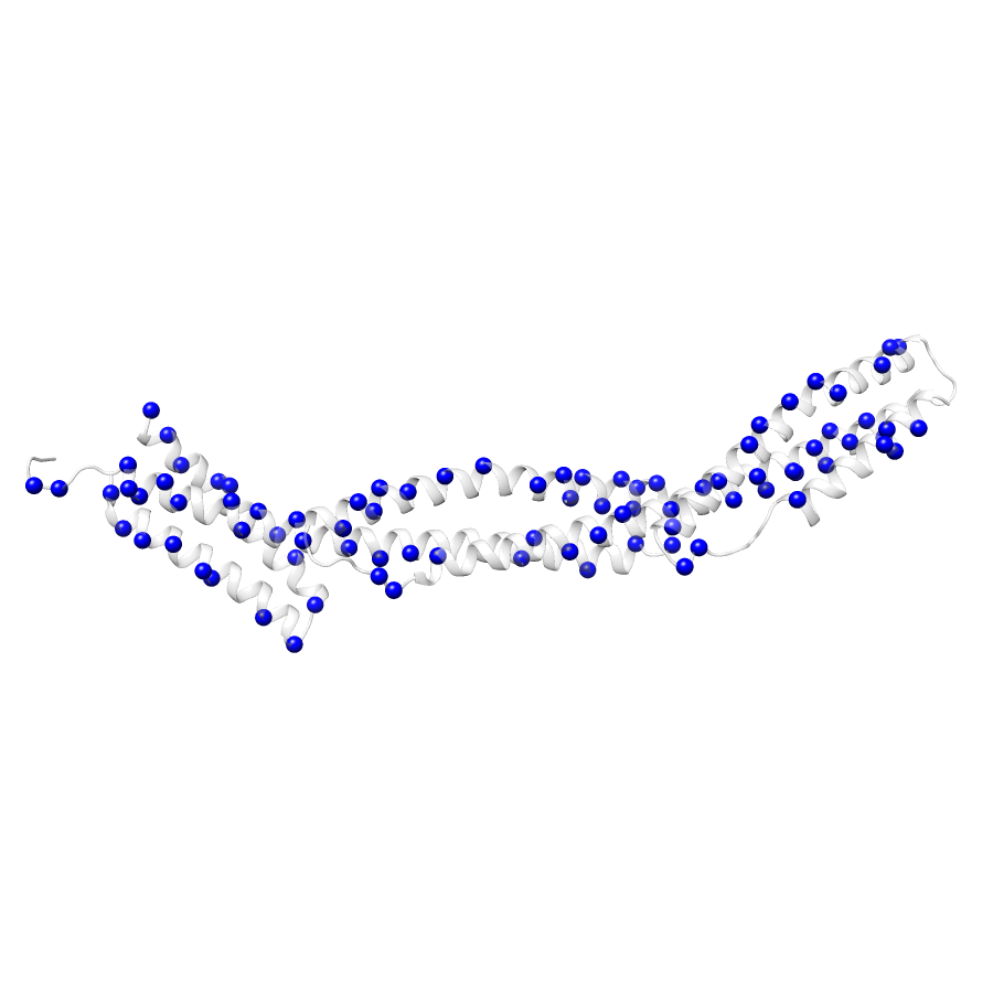
| Number Of Residues | 294 |
| Number Of SNVs | 88 |
| Number Of Permutations | 75195 |
| Optimal Distance Threshold | 13.0 |
| K Statistic | 0.085 |
| p-value | 0.653 |
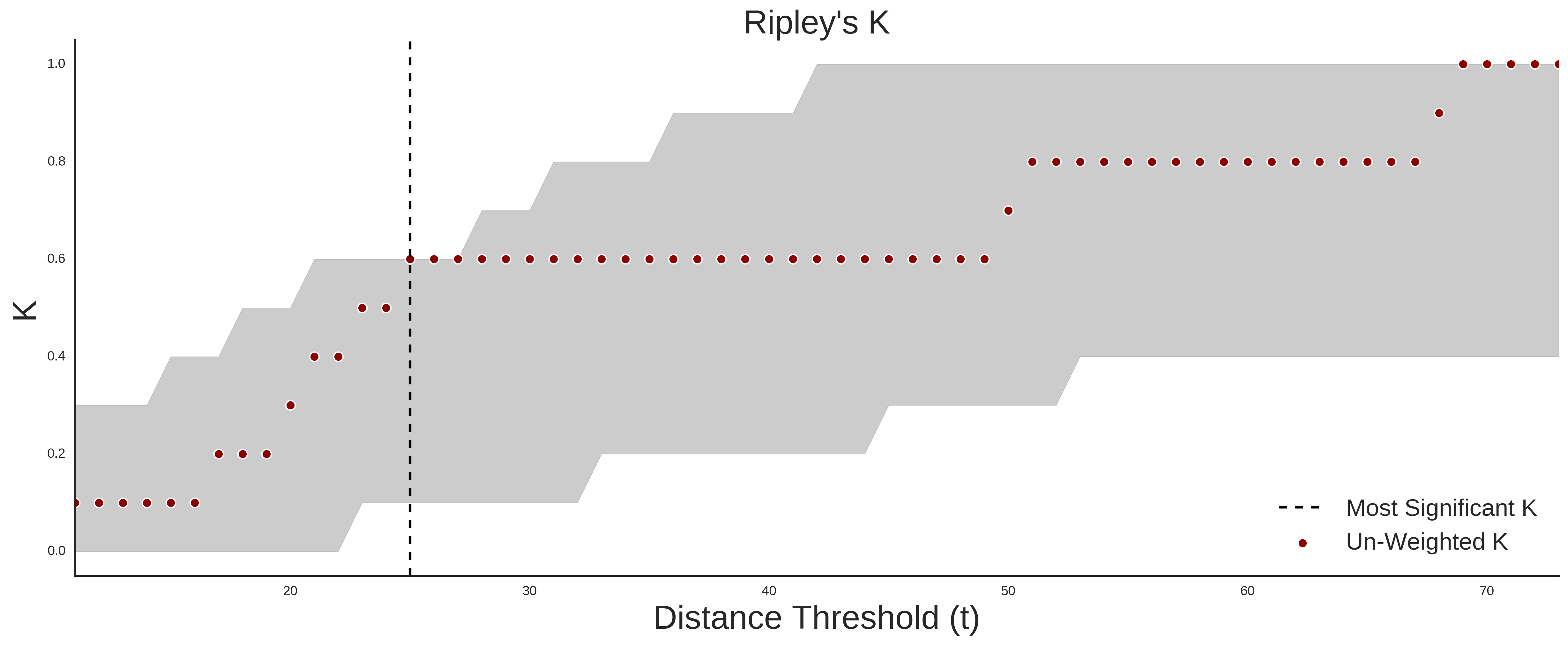
COSMIC
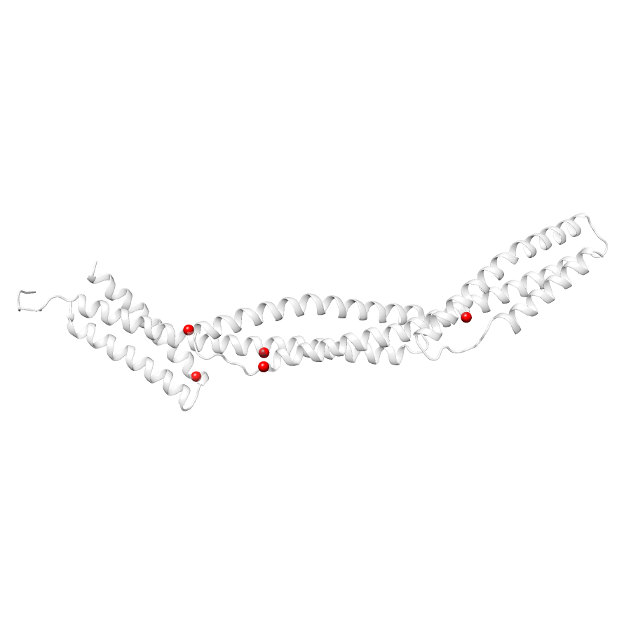
| Number Of Residues | 294 |
| Number Of SNVs | 4 |
| Number Of Permutations | 16 |
| Optimal Distance Threshold | 11.0 |
| K Statistic | 0.167 |
| p-value | 0.685 |
Ripley’s K Analysis Plots
ExACCOSMIC
Variant Set Comparisons
Cosmic vs. ExAC
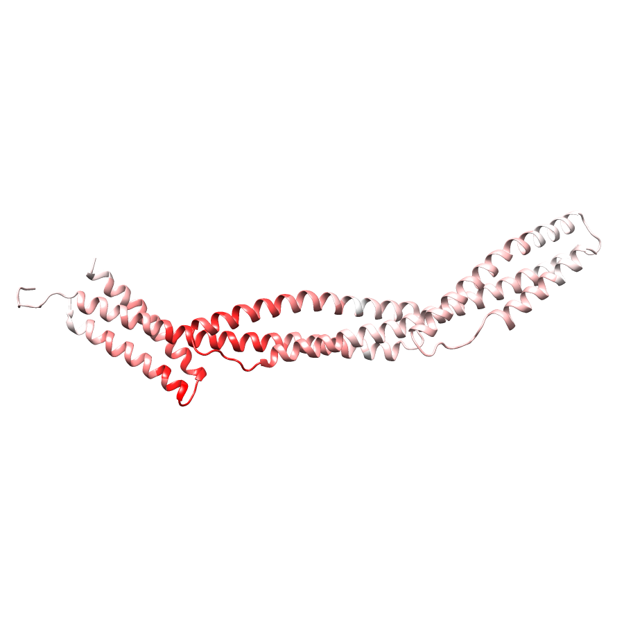
| Number Of ExAC SNVs | 88 |
| Number Of COSMIC SNVs | 4 |
| Optimal Distance Threshold | 25.0 |
| K Statistic | 0.723 |
| p-value | 0.038 |
Pathogenic Proximity Analysis
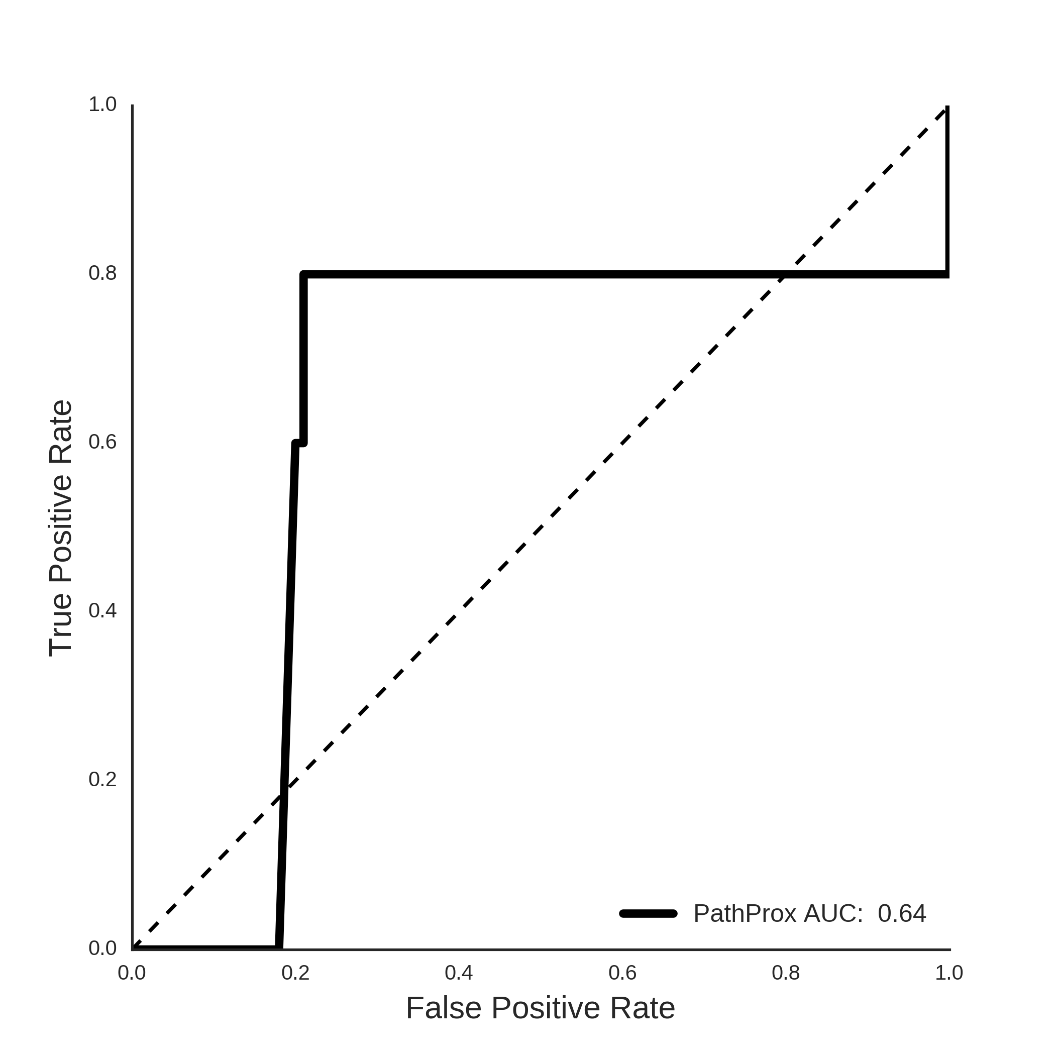
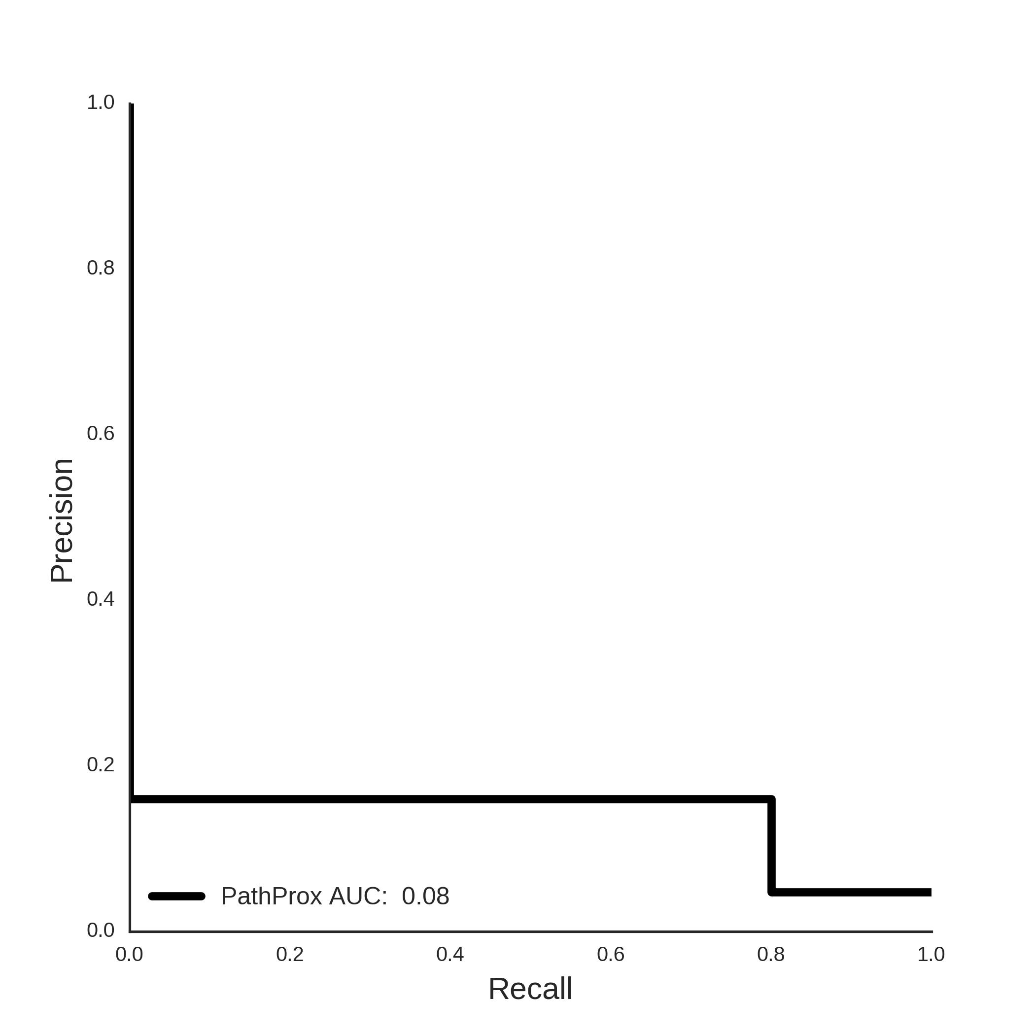
COSMIC PathProx Analysis