General Structure Information
| PDB ID | 1qub |
| HGNC Gene Label(s) | APOH |
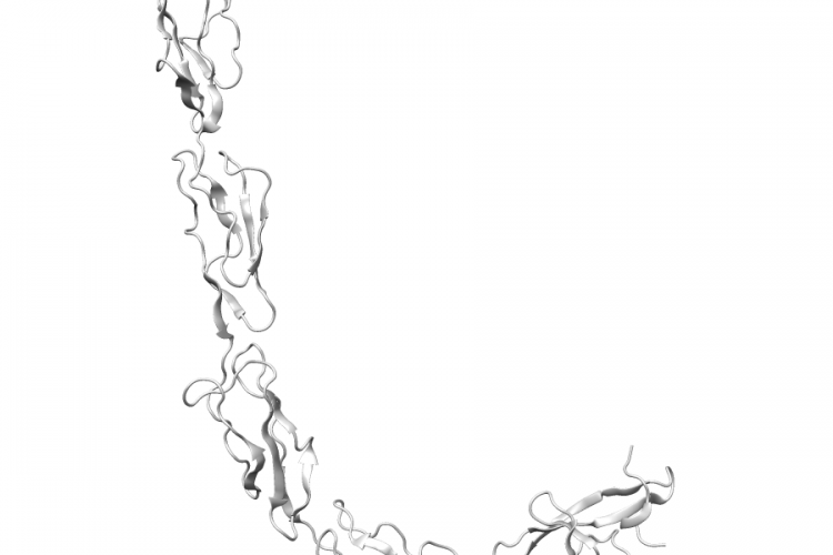
| Structure Name | crystal structure of the glycosylated five-domain human beta2- glycoprotein i purified from blood plasma |
| Resolution | 2.7Å |
| Reference | AUTH B.BOUMA,P.G.DE GROOT,J.M.VAN DEN ELSEN,R.B.RAVELLI,AUTH 2 A.SCHOUTEN,M.J.SIMMELINK,R.H.DERKSEN,J.KROON,P.GROSTITL ADHESION MECHANISM OF HUMAN BETA(2)-GLYCOPROTEIN I TOTITL 2 PHOSPHOLIPIDS BASED ON ITS CRYSTAL STRUCTURE.REF EMBO J. V. 18 5166 1999REFN ISSN 0261-4189PMID 10508150DOI 10.1093/EMBOJ/18.19.5166 |
Variant Set Distributions
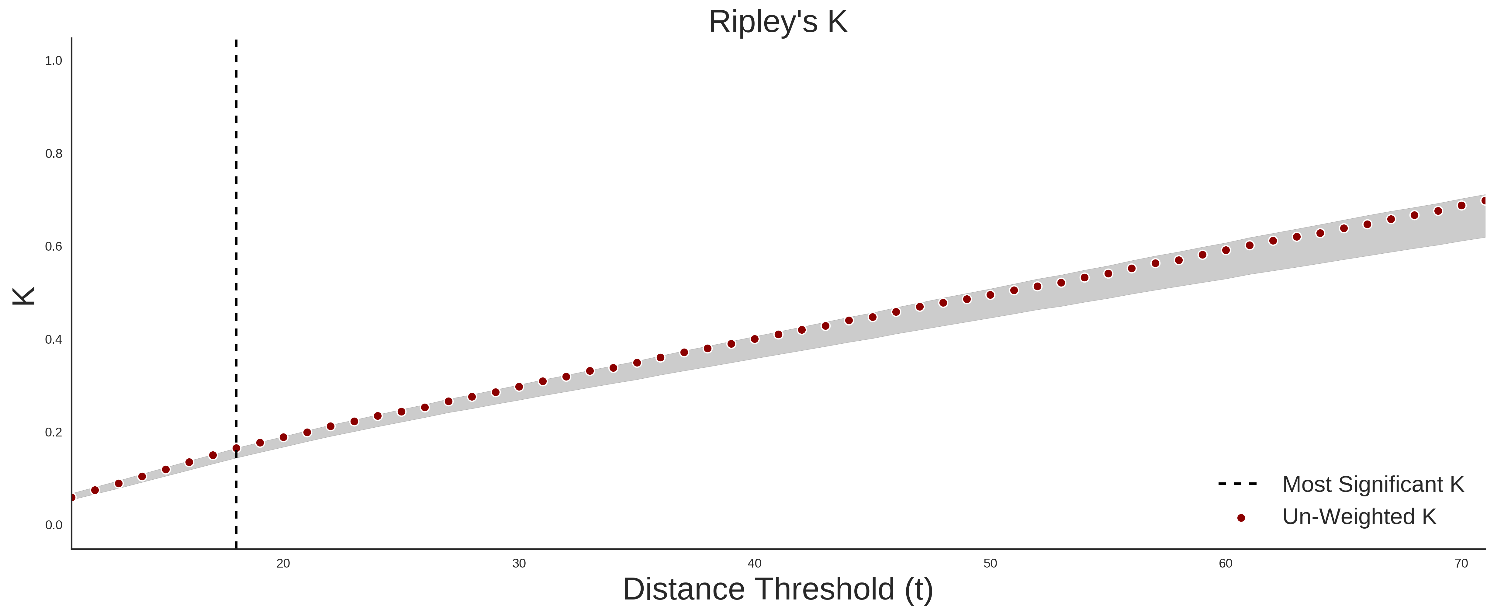
ExAC Variants
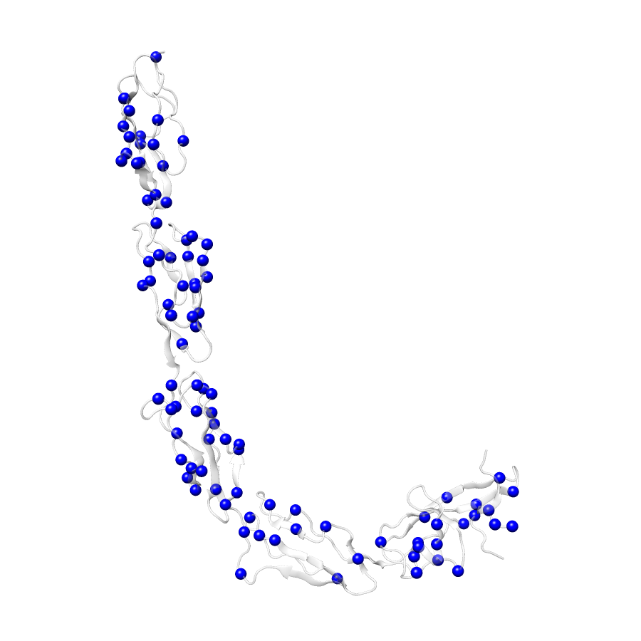
| Number Of Residues | 319 |
| Number Of SNVs | 93 |
| Number Of Permutations | 32902 |
| Optimal Distance Threshold | 18.0 |
| K Statistic | 0.169 |
| p-value | 0.156 |
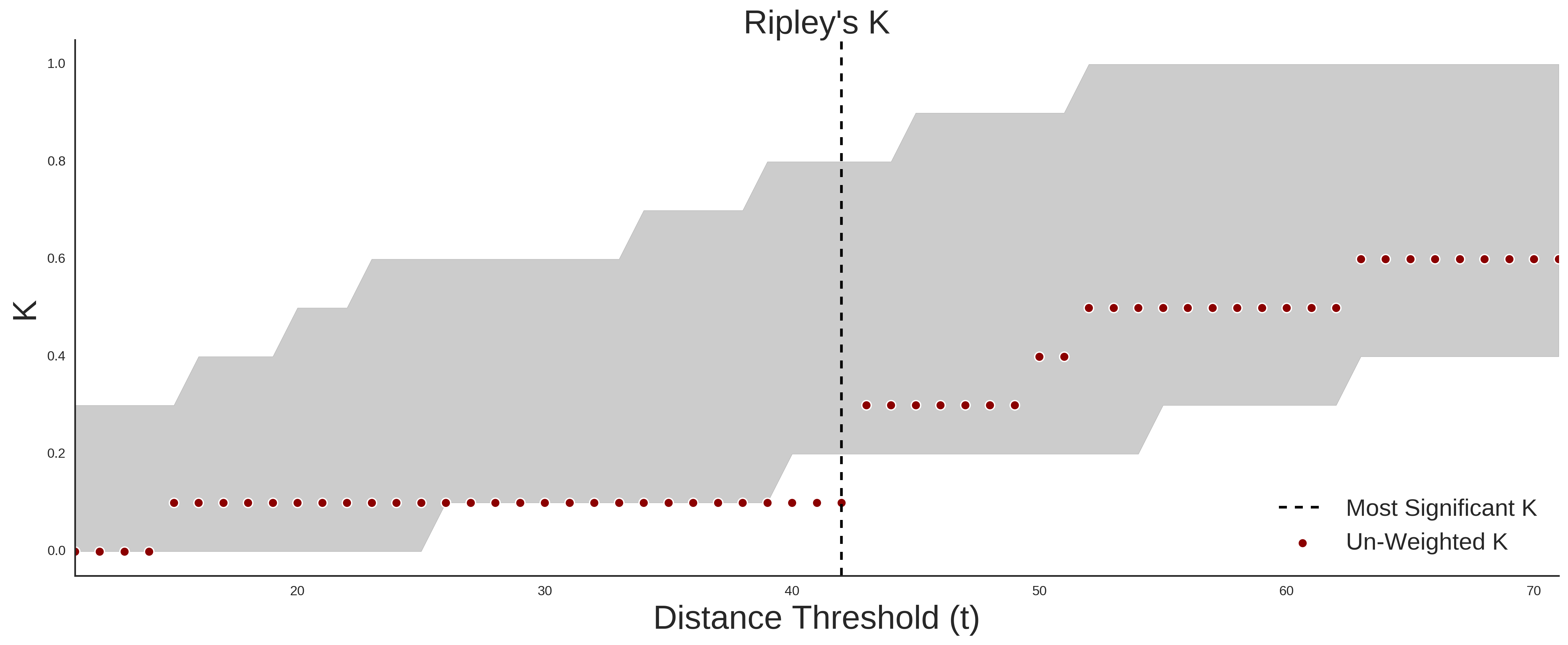
COSMIC
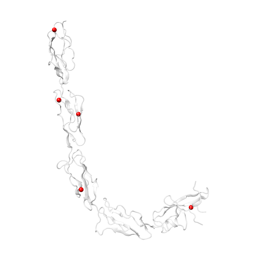
| Number Of Residues | 319 |
| Number Of SNVs | 5 |
| Number Of Permutations | 2535 |
| Optimal Distance Threshold | 42.0 |
| K Statistic | 0.1 |
| p-value | 0.267 |
Ripley’s K Analysis Plots
ExACCOSMIC
Variant Set Comparisons
Cosmic vs. ExAC
| Number Of ExAC SNVs | 93 |
| Number Of COSMIC SNVs | 5 |
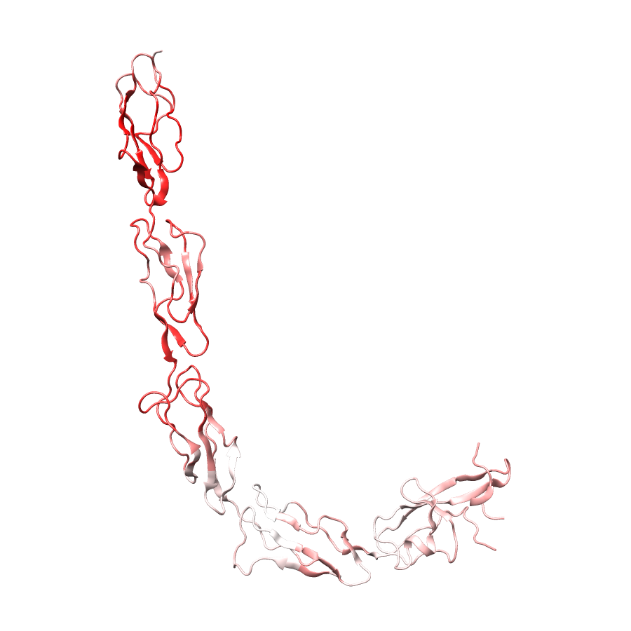
| Optimal Distance Threshold | 42.0 |
| K Statistic | -0.323 |
| p-value | 0.286 |
Pathogenic Proximity Analysis
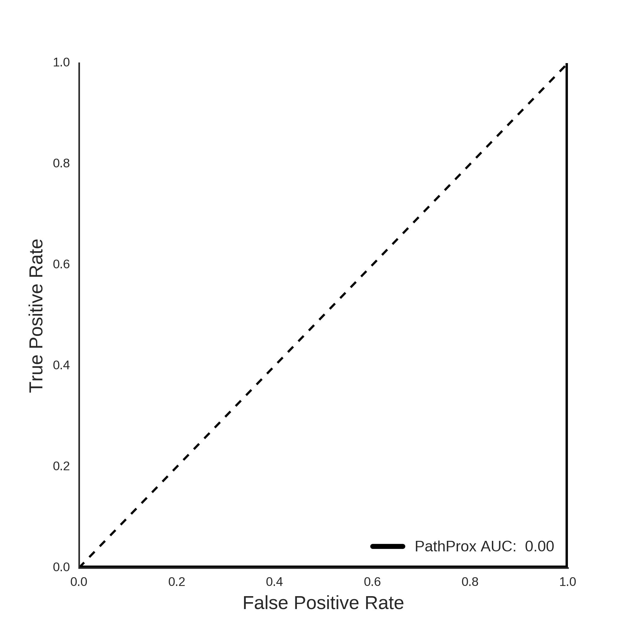
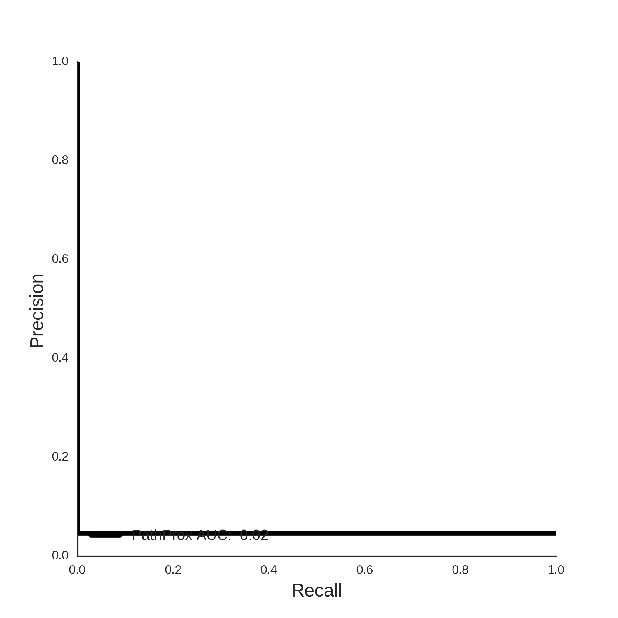
COSMIC PathProx Analysis