General Structure Information
| PDB ID | 4kkd |
| HGNC Gene Label(s) | MASP1 |
| Structure Name | the x-ray crystal structure of mannose-binding lectin-associated serine proteinase-3 reveals the structural basis for enzyme inactivity associated with the 3mc syndrome |
| Resolution | 2.6Å |
| Reference | AUTH T.YONGQING,P.G.WILMANN,S.B.REEVE,T.H.COETZER,A.I.SMITH,AUTH 2 J.C.WHISSTOCK,R.N.PIKE,L.C.WIJEYEWICKREMATITL THE X-RAY CRYSTAL STRUCTURE OF MANNOSE-BINDINGTITL 2 LECTIN-ASSOCIATED SERINE PROTEINASE-3 REVEALS THE STRUCTURALTITL 3 BASIS FOR ENZYME INACTIVITY ASSOCIATED WITH THE CARNEVALE,TITL 4 MINGARELLI, MALPUECH, AND MICHELS (3MC) SYNDROME.REF J.BIOL.CHEM. V. 288 22399 2013REFN ISSN 0021-9258PMID 23792966DOI 10.1074/JBC.M113.483875 |
Variant Set Distributions
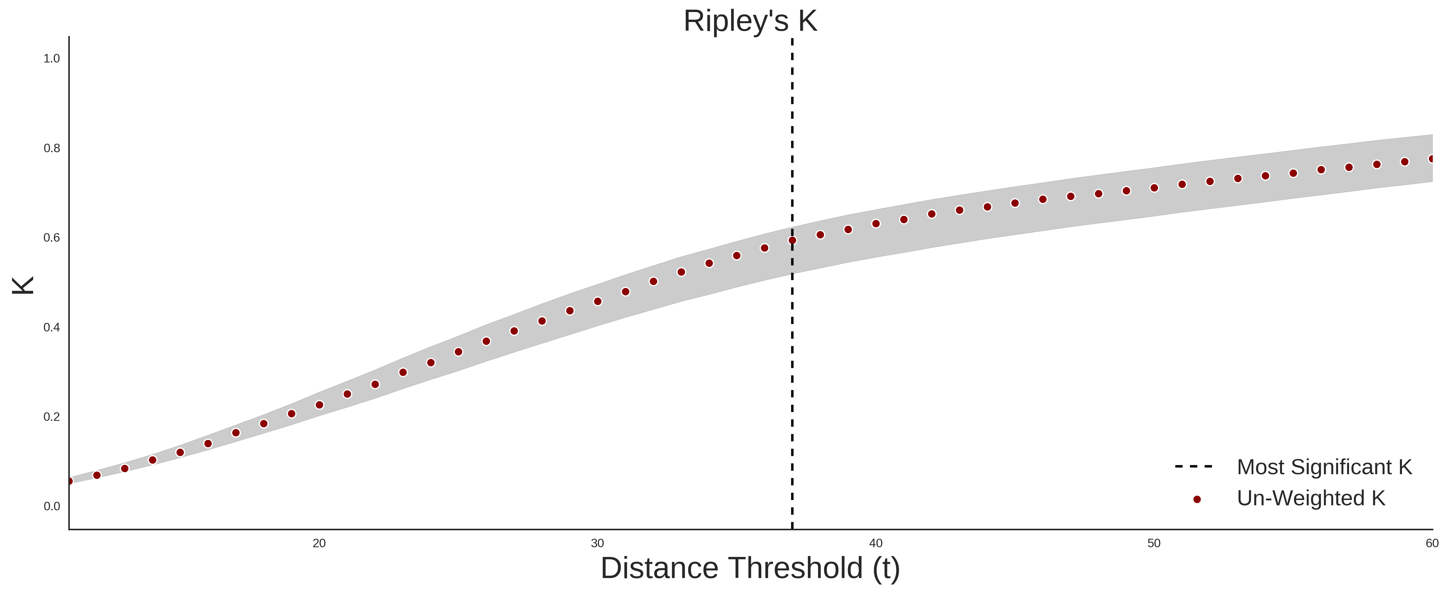
ExAC Variants
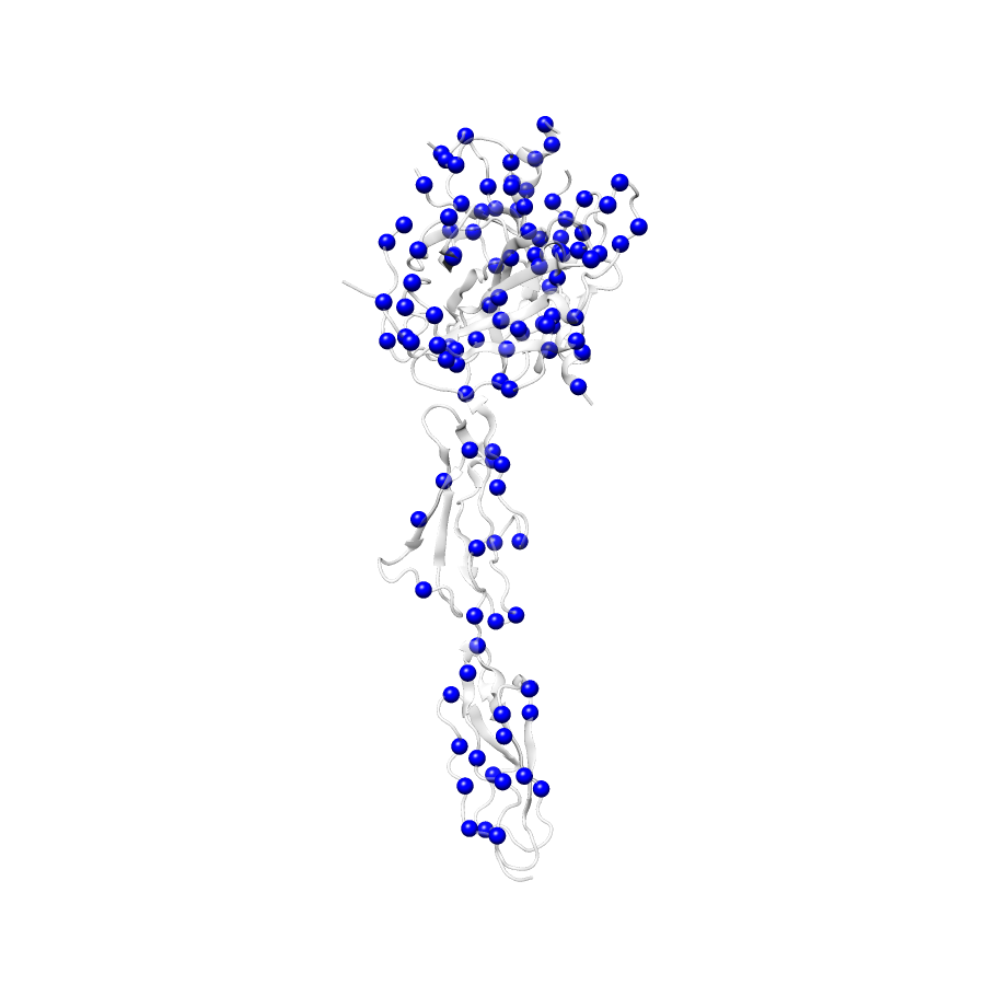
| Number Of Residues | 388 |
| Number Of SNVs | 111 |
| Number Of Permutations | 45526 |
| Optimal Distance Threshold | 37.0 |
| K Statistic | 0.589 |
| p-value | 0.552 |
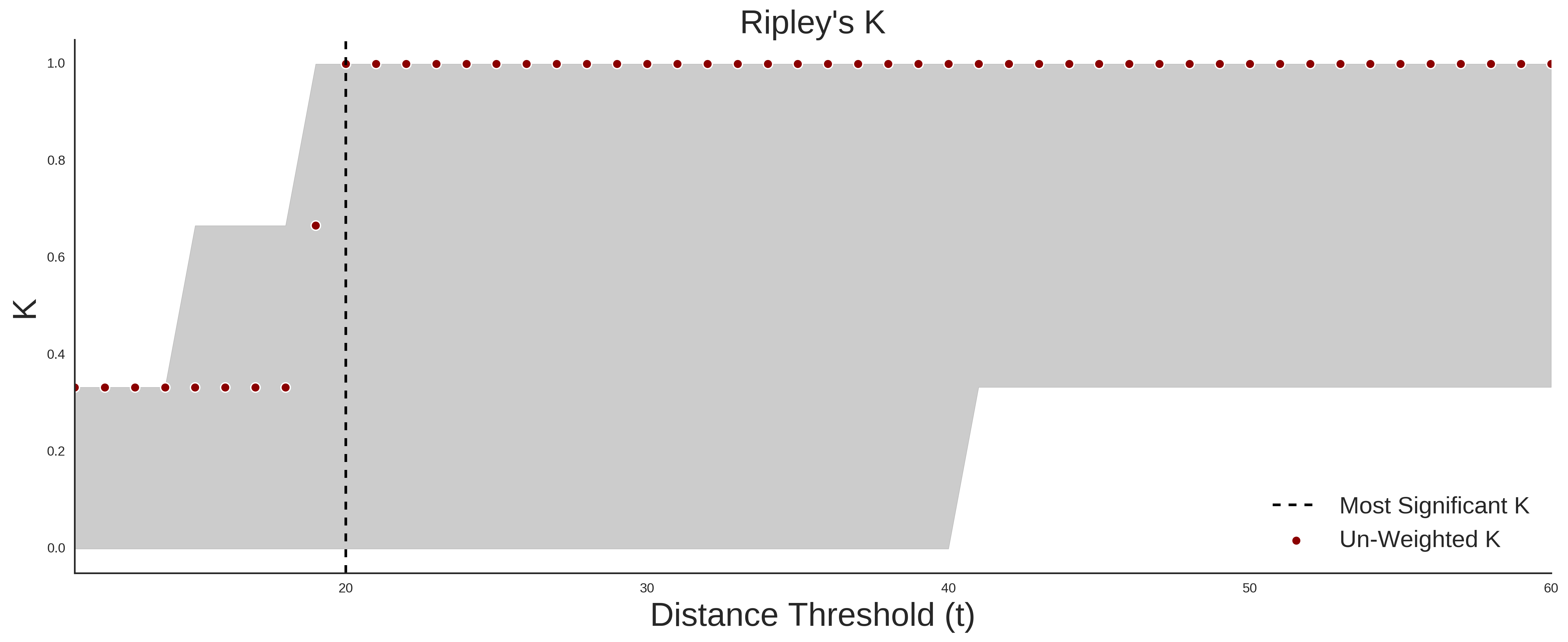
ClinVar
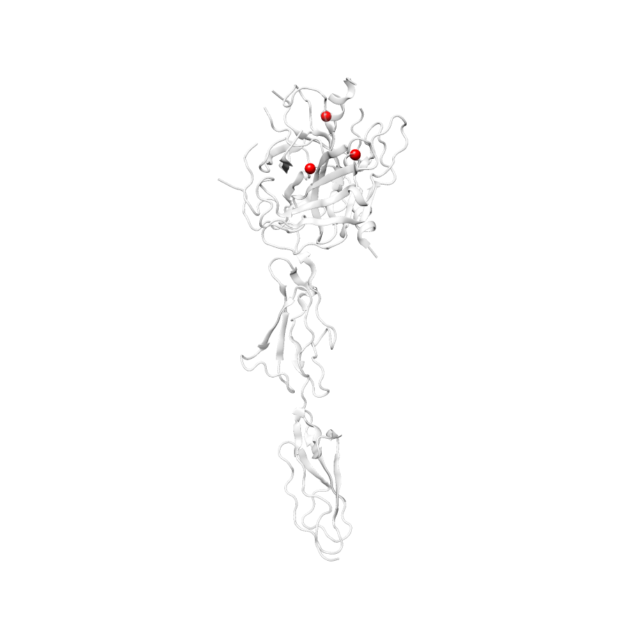
| Number Of Residues | 388 |
| Number Of SNVs | 3 |
| Number Of Permutations | 488 |
| Optimal Distance Threshold | 20.0 |
| K Statistic | 1.0 |
| p-value | 0.092 |
Ripley’s K Analysis Plots
ExACClinVar
Variant Set Comparisons
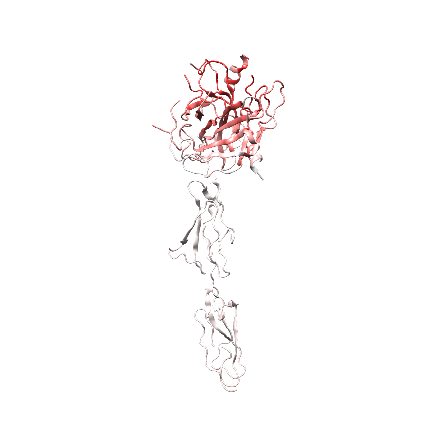
ClinVar vs. ExAC
| Number Of ExAC SNVs | 111 |
| Number Of ClinVar SNVs | 3 |
| Optimal Distance Threshold | 20.0 |
| K Statistic | 0.766 |
| p-value | 0.106 |
Pathogenic Proximity Analysis
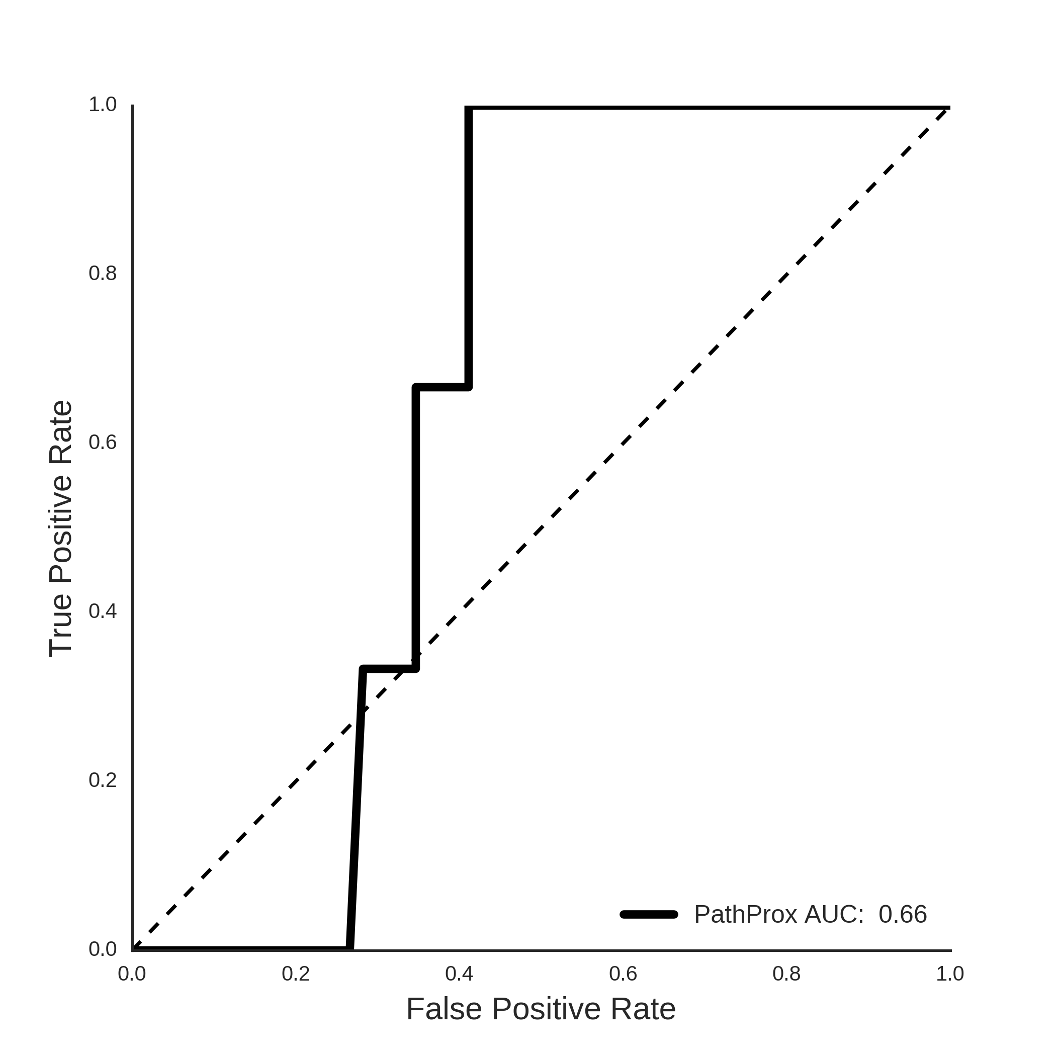
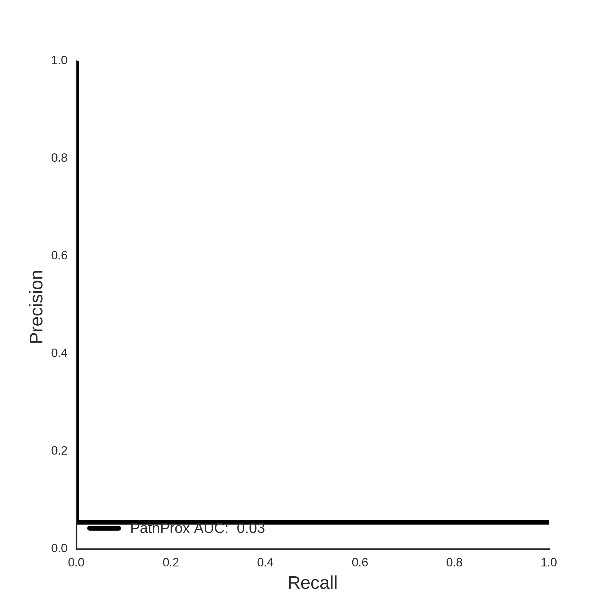
ClinVar PathProx Analysis