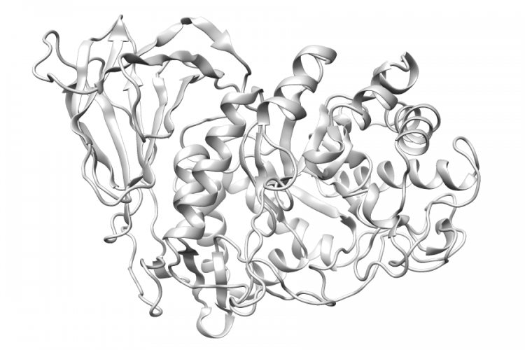
General Structure Information
| PDB ID | 3gxm |
| HGNC Gene Label(s) | GBA |
| Structure Name | crystal structure of acid-beta-glucosidase at ph 4.5, phosphate crystallization condition |
| Resolution | 2.2Å |
| Reference | AUTH R.L.LIEBERMAN,J.A.DAQUINO,D.RINGE,G.A.PETSKOTITL EFFECTS OF PH AND IMINOSUGAR PHARMACOLOGICAL CHAPERONES ONTITL 2 LYSOSOMAL GLYCOSIDASE STRUCTURE AND STABILITY.REF BIOCHEMISTRY V. 48 4816 2009REFN ISSN 0006-2960PMID 19374450DOI 10.1021/BI9002265 |
Variant Set Distributions
ExAC Variants
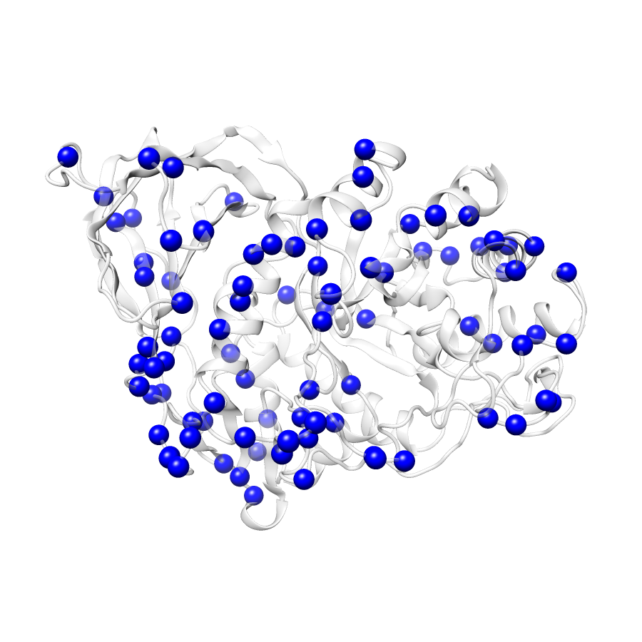
| Number Of Residues | 497 |
| Number Of SNVs | 89 |
| Number Of Permutations | 17945 |
| Optimal Distance Threshold | 6.0 |
| K Statistic | 0.013 |
| p-value | 0.435 |
ClinVar
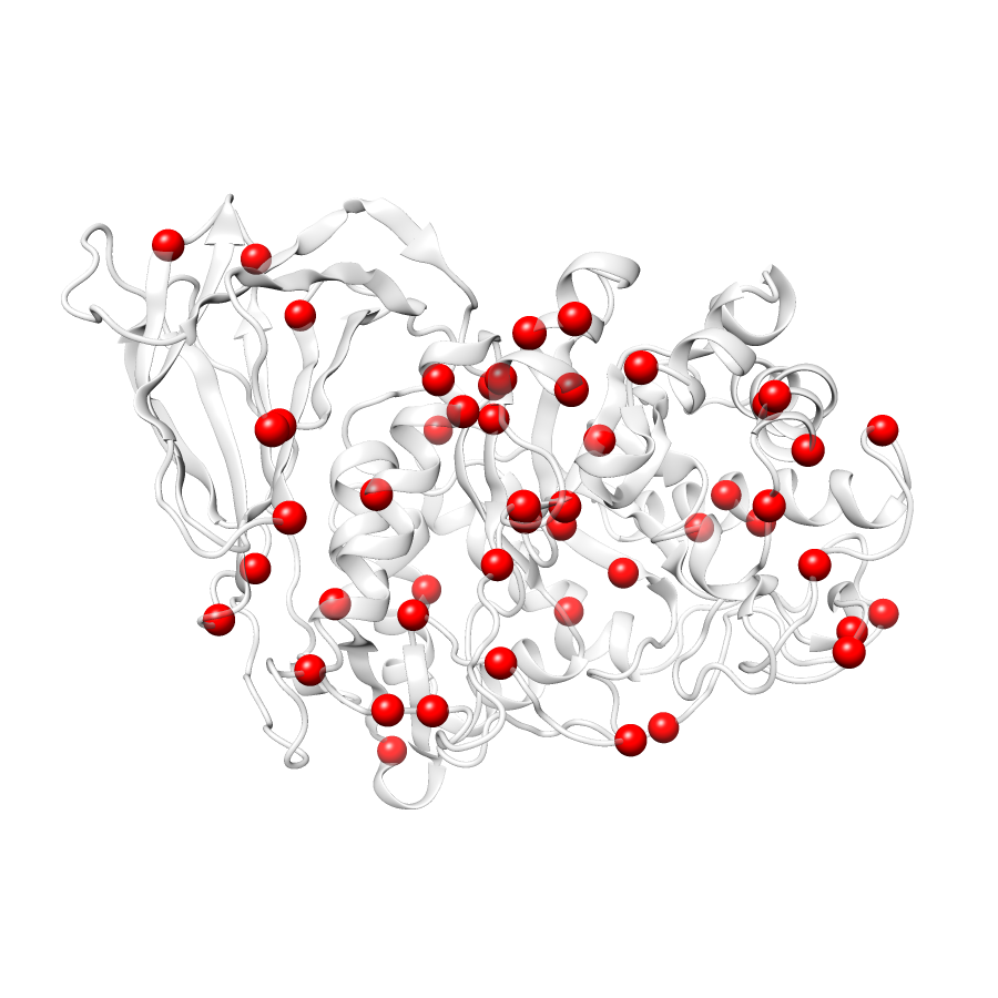
| Number Of Residues | 497 |
| Number Of SNVs | 50 |
| Number Of Permutations | 10163 |
| Optimal Distance Threshold | 8.0 |
| K Statistic | 0.033 |
| p-value | 0.035 |
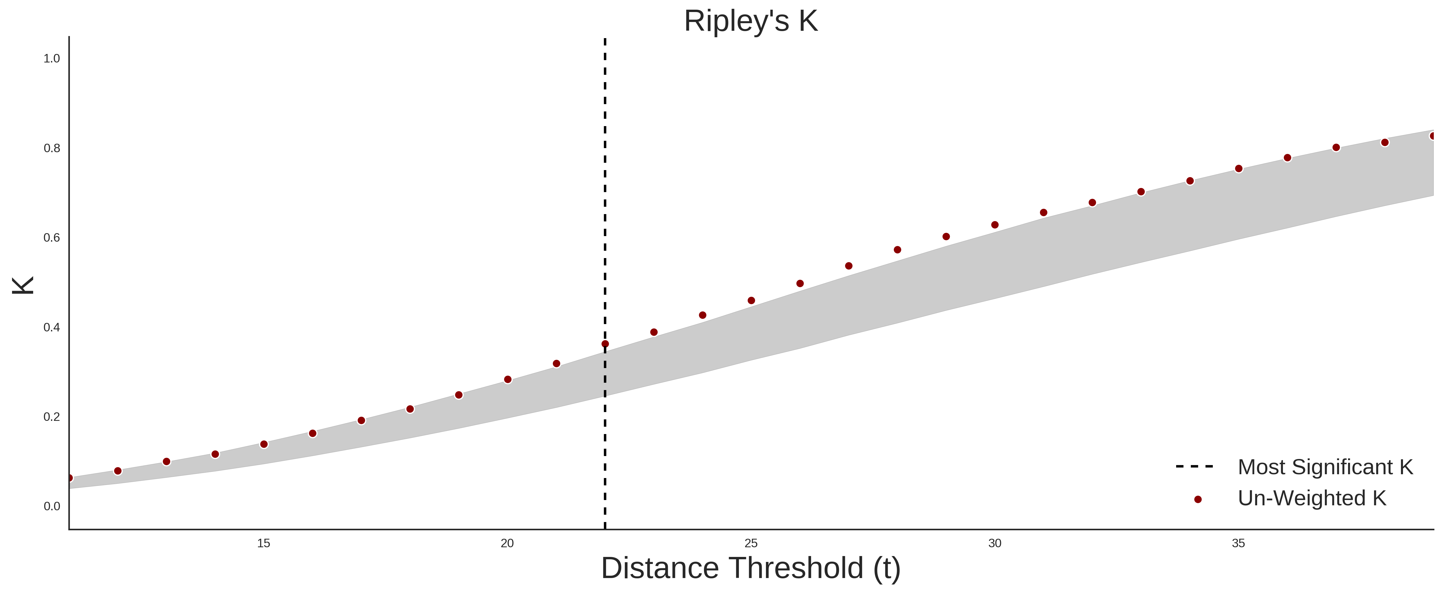
Ripley’s K Analysis Plots
ExACClinVar
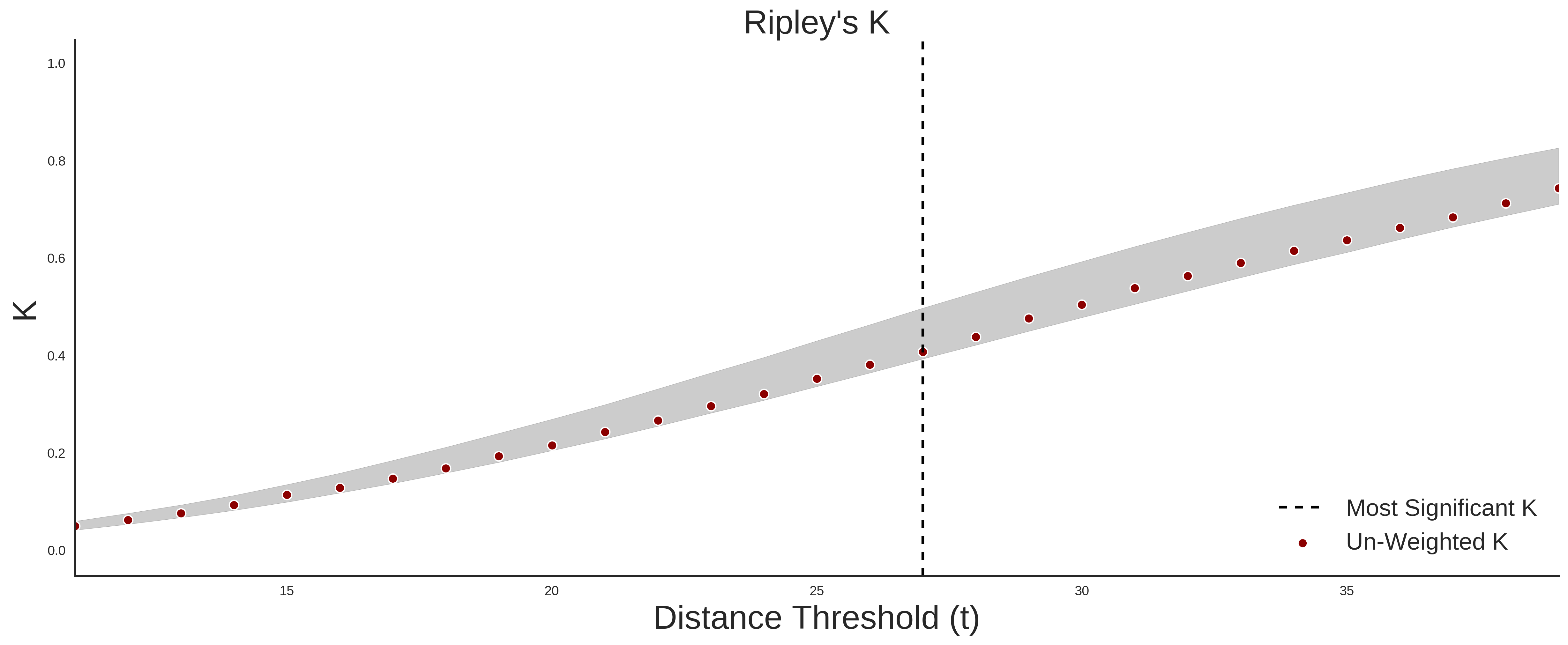
Variant Set Comparisons
ClinVar vs. ExAC
| Number Of ExAC SNVs | 89 |
| Number Of ClinVar SNVs | 48 |
| Optimal Distance Threshold | 22.0 |
| K Statistic | 0.085 |
| p-value | 0.053 |
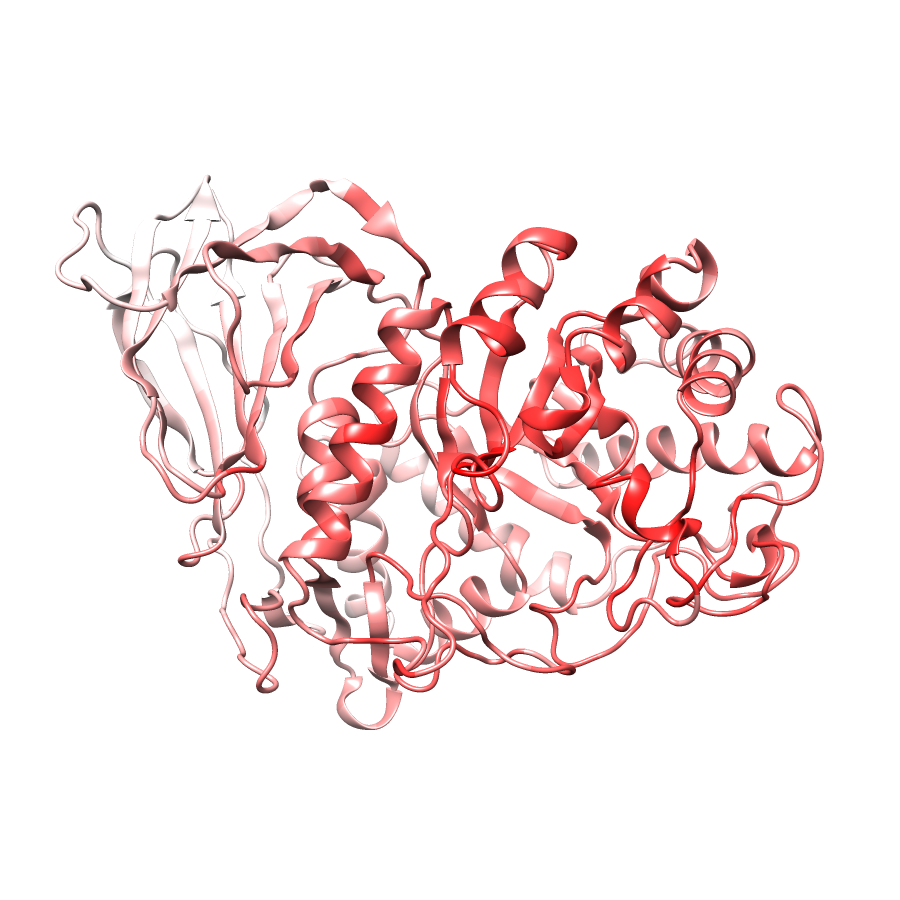
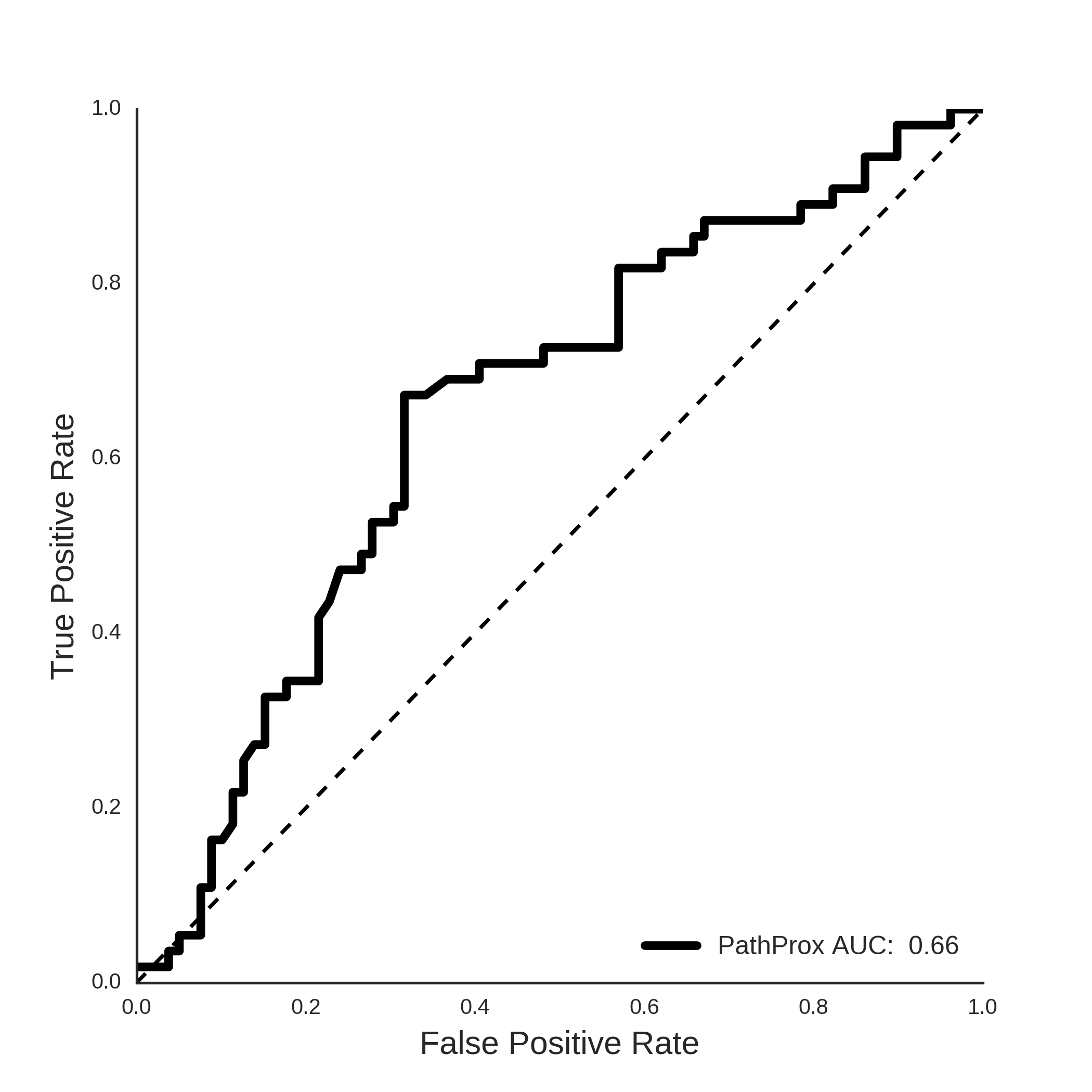
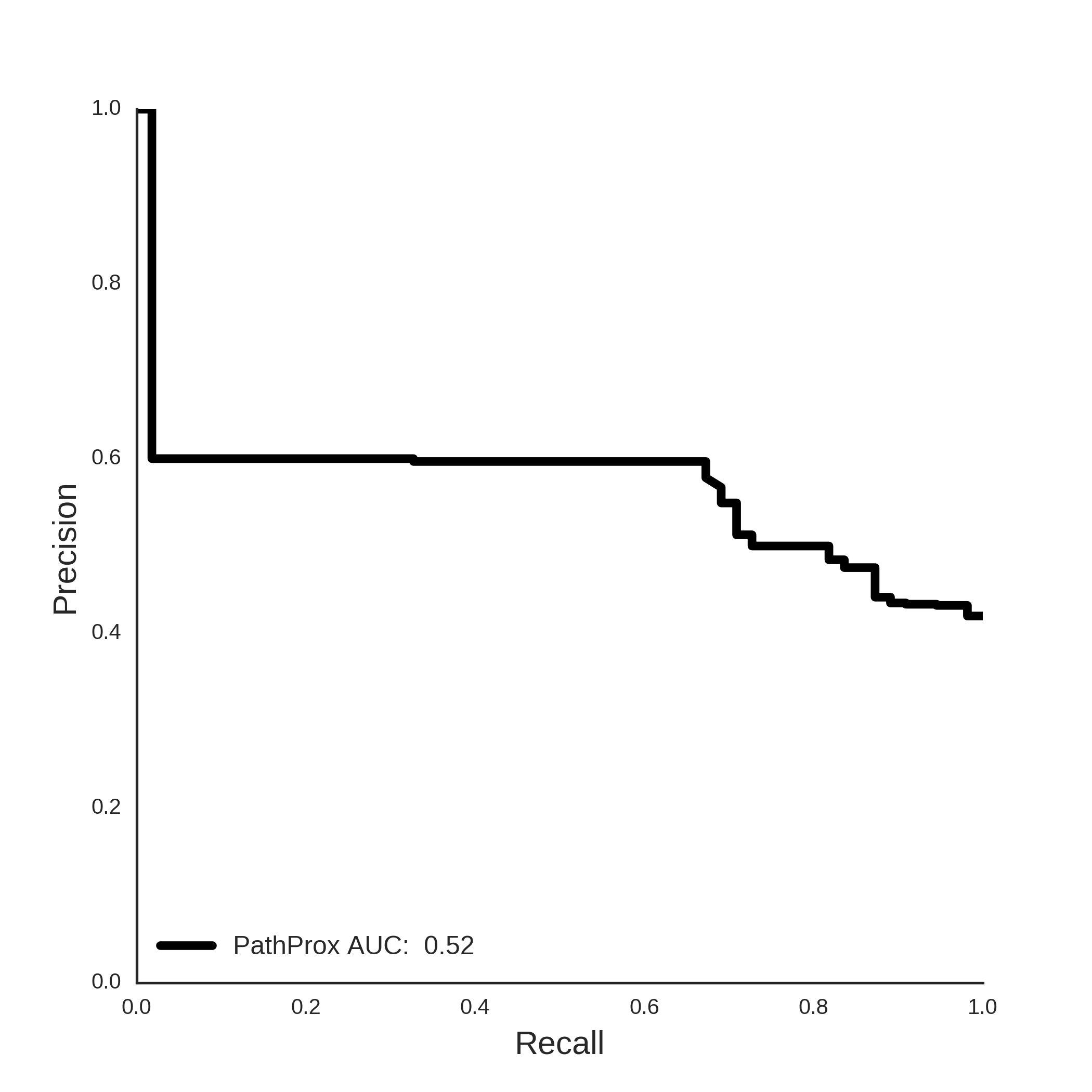
Pathogenic Proximity Analysis
ClinVar PathProx Analysis
Mapped Variants
[+] ExAC Missense Variants
| Position | Variant | AA Change | PDB Chain Position | Structure Coordinates |
|---|---|---|---|---|
| 1:155209863 | rs190207858 | R>C | A:2 | (-25.16, -9.94, 73.08) |
| 1:155209860 | P>S | A:3 | (-19.90, -9.35, 73.86) | |
| 1:155209851 | P>S | A:6 | (-13.10, -14.48, 70.75) | |
| 1:155209848 | rs142761046 | K>E | A:7 | (-12.00, -18.39, 66.78) |
| 1:155209844 | rs543005460 | S>I | A:8 | (-7.73, -17.74, 66.47) |
| 1:155209839 | G>S | A:10 | (-3.28, -17.64, 63.05) | |
| 1:155209836 | Y>H | A:11 | (-4.05, -15.66, 60.05) | |
| 1:155209820 | C>S | A:16 | (-16.11, -12.03, 65.95) | |
| 1:155209811 | N>S | A:19 | (-24.53, -18.03, 66.03) | |
| 1:155209799 | rs145888253 | C>Y | A:23 | (-24.07, -11.50, 67.52) |
| 1:155209778 | T>I | A:30 | (-26.29, 7.25, 74.26) | |
| 1:155209769 | A>D | A:33 | (-22.45, 16.11, 78.13) | |
| 1:155209760 | T>N | A:36 | (-18.00, 14.34, 78.14) | |
| 1:155209761 | T>A | A:36 | (-18.00, 14.34, 78.14) | |
| 1:155209753 | S>R | A:38 | (-17.39, 9.08, 73.44) | |
| 1:155209751 | R>H | A:39 | (-12.48, 6.28, 74.06) | |
| 1:155209752 | rs146774384 | R>C | A:39 | (-12.48, 6.28, 74.06) |
| 1:155209739 | rs1141811 | T>I | A:43 | (-7.26, -3.65, 69.77) |
| 1:155209736 | R>H | A:44 | (-2.57, -6.76, 66.83) | |
| 1:155209737 | rs1141812 | R>C | A:44 | (-2.57, -6.76, 66.83) |
| 1:155209730 | rs77829017 | G>E | A:46 | (-9.19, -7.66, 70.96) |
| 1:155209727 | rs144173415 | R>Q | A:47 | (-9.76, -3.88, 73.79) |
| 1:155209725 | rs1141814 | R>W | A:48 | (-12.68, -5.92, 68.31) |
| 1:155209720 | M>I | A:49 | (-16.36, -1.03, 68.22) | |
| 1:155209701 | I>V | A:56 | (-13.36, 17.66, 77.17) | |
| 1:155209689 | rs1141818 | H>N | A:60 | (-2.30, 20.72, 77.33) |
| 1:155209490 | M>R | A:85 | (-26.03, -7.68, 47.59) | |
| 1:155209491 | M>V | A:85 | (-26.03, -7.68, 47.59) | |
| 1:155209478 | A>V | A:89 | (-23.34, -12.54, 49.16) | |
| 1:155209448 | COSM252999 COSM252517 |
P>H | A:99 | (-32.08, -16.71, 62.77) |
| 1:155209449 | P>A | A:99 | (-32.08, -16.71, 62.77) | |
| 1:155209439 | rs374003673 | N>S | A:102 | (-31.80, -11.64, 60.78) |
| 1:155209424 | S>L | A:107 | (-24.23, -6.47, 61.77) | |
| 1:155209407 | G>R | A:113 | (-24.59, -1.89, 64.30) | |
| 1:155208060 | R>H | A:170 | (-28.39, 5.67, 59.07) | |
| 1:155208061 | R>C | A:170 | (-28.39, 5.67, 59.07) | |
| 1:155208055 | rs188760929 | V>I | A:172 | (-30.60, 2.82, 52.02) |
| 1:155208049 | L>F | A:174 | (-29.90, 1.41, 46.05) | |
| 1:155208019 | rs61748906 | W>R | A:184 | (-27.62, -9.18, 21.52) |
| 1:155208005 | rs381418 | N>K | A:188 | (-16.19, -10.29, 21.74) |
| 1:155208006 | rs364897 | N>S | A:188 | (-16.19, -10.29, 21.74) |
| 1:155208000 | A>V | A:190 | (-18.32, -12.39, 24.74) | |
| 1:155207997 | rs381427 | V>G | A:191 | (-19.34, -12.50, 28.68) |
| 1:155207983 | rs1064644 | S>P | A:196 | (-16.73, -3.27, 22.88) |
| 1:155207977 | K>E | A:198 | (-21.64, -4.13, 17.63) | |
| 1:155207965 | rs398123534 COSM529328 CM940807 |
G>R | A:202 | (-26.42, 6.18, 18.42) |
| 1:155207946 | T>N | A:208 | (-30.37, -1.44, 23.27) | |
| 1:155207935 | rs121908300 | Y>H | A:212 | (-29.97, -3.74, 30.14) |
| 1:155207932 | rs381737 | F>I | A:213 | (-28.67, 1.18, 32.26) |
| 1:155207249 | rs367968666 CM000163 |
H>Q | A:255 | (-16.95, 4.40, 23.61) |
| 1:155207244 | rs78973108 | R>Q | A:257 | (-16.69, 11.43, 25.11) |
| 1:155207229 | rs140955685 | R>H | A:262 | (-25.10, 10.51, 20.91) |
| 1:155207230 | R>C | A:262 | (-25.10, 10.51, 20.91) | |
| 1:155207218 | P>A | A:266 | (-30.02, 11.04, 26.58) | |
| 1:155207214 | T>I | A:267 | (-32.18, 7.34, 27.61) | |
| 1:155207209 | rs368425393 | A>T | A:269 | (-33.79, 11.85, 30.85) |
| 1:155207192 | COSM4022964 | H>Q | A:274 | (-35.85, 12.10, 36.06) |
| 1:155207193 | H>R | A:274 | (-35.85, 12.10, 36.06) | |
| 1:155207190 | rs535896234 | N>S | A:275 | (-34.20, 11.89, 39.67) |
| 1:155207160 | rs79696831 | R>H | A:285 | (-6.24, 6.17, 42.87) |
| 1:155207144 | rs142662866 | H>Q | A:290 | (-7.14, 11.13, 30.63) |
| 1:155206260 | V>I | A:295 | (-16.01, 12.81, 30.07) | |
| 1:155206227 | rs181720335 | H>Y | A:306 | (-23.53, 13.55, 40.57) |
| 1:155206217 | A>G | A:309 | (-16.25, 6.21, 40.38) | |
| 1:155206200 | rs398123526 CM950565 |
D>N | A:315 | (-4.85, 2.78, 45.15) |
| 1:155206184 | A>G | A:320 | (-3.93, 10.70, 46.09) | |
| 1:155206175 | rs76539814 | T>I | A:323 | (-5.32, 10.90, 41.36) |
| 1:155206167 | rs2230288 | E>K | A:326 | (-5.02, 17.39, 40.07) |
| 1:155206157 | rs1064648 | R>H | A:329 | (-6.95, 21.63, 40.37) |
| 1:155206158 | R>C | A:329 | (-6.95, 21.63, 40.37) | |
| 1:155206122 | A>T | A:341 | (-13.78, -0.52, 48.52) | |
| 1:155206104 | F>L | A:347 | (2.38, -13.38, 44.94) | |
| 1:155206101 | W>G | A:348 | (2.76, -13.08, 49.24) | |
| 1:155206093 | Q>H | A:350 | (-5.07, -17.85, 51.58) | |
| 1:155206085 | rs11558184 | R>Q | A:353 | (-4.74, -13.79, 56.14) |
| 1:155206076 | S>F | A:356 | (-6.33, -9.81, 59.37) | |
| 1:155206060 | rs149487315 | M>I | A:361 | (-4.46, -1.59, 59.50) |
| 1:155206052 | rs121908307 | S>T | A:364 | (-8.80, 2.55, 57.90) |
| 1:155206049 | H>P | A:365 | (-4.44, 4.40, 55.34) | |
| 1:155206041 | I>V | A:368 | (-8.42, 8.30, 56.51) | |
| 1:155206037 | rs75548401 | T>M | A:369 | (-6.48, 8.84, 52.22) |
| 1:155205634 | rs76763715 | N>S | A:370 | (-10.27, 8.51, 50.22) |
| 1:155205614 | rs121908311 | G>S | A:377 | (-17.84, 7.59, 49.72) |
| 1:155205596 | rs187143994 | L>I | A:383 | (-21.77, -7.70, 52.01) |
| 1:155205592 | A>V | A:384 | (-20.16, -12.04, 52.07) | |
| 1:155205589 | L>P | A:385 | (-17.23, -14.21, 50.77) | |
| 1:155205581 | rs149171124 | E>K | A:388 | (-12.98, -20.92, 50.23) |
| 1:155205568 | N>S | A:392 | (-15.46, -16.53, 41.75) | |
| 1:155205563 | rs80356769 | V>L | A:394 | (-12.97, -17.10, 37.48) |
| 1:155205559 | R>H | A:395 | (-8.26, -17.51, 40.96) | |
| 1:155205516 | rs78802049 | D>E | A:409 | (-25.46, -21.66, 61.25) |
| 1:155205518 | rs1064651 | D>H | A:409 | (-25.46, -21.66, 61.25) |
| 1:155205514 | T>M | A:410 | (-21.22, -19.88, 61.75) | |
| 1:155205062 | A>T | A:438 | (-1.01, 18.69, 63.40) | |
| 1:155205055 | rs144389406 | Q>P | A:440 | (4.35, 17.19, 66.16) |
| 1:155205047 | rs75671029 | D>N | A:443 | (3.28, 7.96, 62.50) |
| 1:155205038 | A>T | A:446 | (-6.51, 11.48, 61.13) | |
| 1:155205016 | D>V | A:453 | (-25.75, 17.68, 65.23) | |
| 1:155205005 | COSM1498627 | V>I | A:457 | (-16.40, 12.69, 64.41) |
| 1:155204986 | rs80356772 | R>H | A:463 | (-0.66, 2.20, 59.95) |
[+] ClinVar Missense Variants
| Position | Variant | AA Change | PDB Chain Position | Structure Coordinates |
|---|---|---|---|---|
| 1:155209824 | rs121908302 | V>L | A:15 | (-12.70, -13.49, 64.31) |
| 1:155209730 | rs77829017 | G>E | A:46 | (-9.19, -7.66, 70.96) |
| 1:155209725 | rs1141814 | R>W | A:48 | (-12.68, -5.92, 68.31) |
| 1:155209507 | rs121908312 | K>N | A:79 | (-24.12, 9.77, 50.26) |
| 1:155209430 | rs794727708 | L>R | A:105 | (-28.68, -8.27, 57.64) |
| 1:155208420 | rs79653797 | R>Q | A:120 | (-21.83, -1.92, 43.80) |
| 1:155208421 | rs397515515 | R>W | A:120 | (-21.83, -1.92, 43.80) |
| 1:155208415 | rs121908299 | P>S | A:122 | (-24.76, -6.18, 40.73) |
| 1:155208387 | rs80356763 | R>L | A:131 | (-25.75, -18.11, 36.12) |
| 1:155208388 | rs398123530 | R>C | A:131 | (-25.75, -18.11, 36.12) |
| 1:155208361 | rs147138516 | D>H | A:140 | (-31.33, -10.08, 17.80) |
| 1:155208310 | rs121908297 | K>Q | A:157 | (-29.79, -9.66, 44.62) |
| 1:155208061 | rs398123532 | R>C | A:170 | (-28.39, 5.67, 59.07) |
| 1:155208019 | rs61748906 | W>R | A:184 | (-27.62, -9.18, 21.52) |
| 1:155208006 | rs364897 | N>S | A:188 | (-16.19, -10.29, 21.74) |
| 1:155208004 | rs786200979 | NG>RG | A:189 | (-19.42, -12.40, 21.60) |
| 1:155207983 | rs1064644 | S>P | A:196 | (-16.73, -3.27, 22.88) |
| 1:155207965 | rs409652 | G>R | A:202 | (-26.42, 6.18, 18.42) |
| 1:155207935 | rs121908300 | Y>H | A:212 | (-29.97, -3.74, 30.14) |
| 1:155207932 | rs381737 | F>I | A:213 | (-28.67, 1.18, 32.26) |
| 1:155207367 | rs74500255 | F>Y | A:216 | (-31.89, -2.73, 36.54) |
| 1:155207368 | rs121908303 | F>V | A:216 | (-31.89, -2.73, 36.54) |
| 1:155207261 | rs121908313 | F>L | A:251 | (-16.64, 1.65, 27.39) |
| 1:155207249 | rs367968666 | H>Q | A:255 | (-16.95, 4.40, 23.61) |
| 1:155207244 | rs78973108 | R>Q | A:257 | (-16.69, 11.43, 25.11) |
| 1:155207235 | rs794727908 | I>T | A:260 | (-22.67, 9.47, 28.59) |
| 1:155207148 | rs121908298 | P>L | A:289 | (-6.64, 13.11, 35.83) |
| 1:155206217 | rs78396650 | A>V | A:309 | (-16.25, 6.21, 40.38) |
| 1:155206211 | rs78198234 | H>R | A:311 | (-12.65, 0.69, 41.75) |
| 1:155206207 | rs121908304 | W>C | A:312 | (-10.13, 1.56, 48.07) |
| 1:155206200 | rs398123526 | D>N | A:315 | (-4.85, 2.78, 45.15) |
| 1:155206175 | rs76539814 | T>I | A:323 | (-5.32, 10.90, 41.36) |
| 1:155206170 | rs121908305 | G>R | A:325 | (-6.56, 15.84, 43.83) |
| 1:155206167 | rs2230288 | E>K | A:326 | (-5.02, 17.39, 40.07) |
| 1:155206119 | rs121908306 | C>G | A:342 | (-11.28, -3.65, 47.72) |
| 1:155206089 | rs398123527 | V>L | A:352 | (-9.73, -15.60, 53.35) |
| 1:155206086 | rs121908308 | R>G | A:353 | (-4.74, -13.79, 56.14) |
| 1:155206052 | rs121908307 | S>T | A:364 | (-8.80, 2.55, 57.90) |
| 1:155205634 | rs76763715 | N>S | A:370 | (-10.27, 8.51, 50.22) |
| 1:155205632 | rs121908314 | L>V | A:371 | (-12.14, 11.34, 54.01) |
| 1:155205620 | rs398123528 | V>L | A:375 | (-14.01, 10.89, 48.82) |
| 1:155205614 | rs121908311 | G>S | A:377 | (-17.84, 7.59, 49.72) |
| 1:155205563 | rs80356769 | V>L | A:394 | (-12.97, -17.10, 37.48) |
| 1:155205551 | rs121908310 | V>F | A:398 | (-10.98, -10.94, 47.42) |
| 1:155205541 | rs74598136 | P>L | A:401 | (-15.95, -8.11, 56.00) |
| 1:155205517 | rs77369218 | D>V | A:409 | (-25.46, -21.66, 61.25) |
| 1:155205518 | rs1064651 | D>H | A:409 | (-25.46, -21.66, 61.25) |
| 1:155205499 | rs121908295 | P>R | A:415 | (-10.82, -7.20, 61.52) |
| 1:155205047 | rs75671029 | D>N | A:443 | (3.28, 7.96, 62.50) |
| 1:155205043 | rs421016 | L>R | A:444 | (-1.30, 8.93, 63.92) |
| 1:155205008 | rs368060 | A>P | A:456 | (-17.63, 15.78, 66.94) |
| 1:155204986 | rs80356772 | R>H | A:463 | (-0.66, 2.20, 59.95) |
| 1:155204987 | rs80356771 | R>C | A:463 | (-0.66, 2.20, 59.95) |
| 1:155204848 | rs121908301 | G>S | A:478 | (-11.03, 21.57, 74.88) |
| 1:155204793 | rs75822236 | R>H | A:496 | (-18.52, 21.90, 69.19) |