General Structure Information
| PDB ID | 2k1m |
| HGNC Gene Label(s) | MYBPC3 |
| Structure Name | 3d nmr structure of domain cc0 of cardiac myosin binding protein c (mybpc) |
| Resolution | -1.0Å |
| Reference | AUTH J.RATTI,M.GAUTEL,M.PFUHLTITL 3D NMR STRUCTURE OF DOMAIN CC0 OF CARDIAC MYOSINTITL 2 BINDING PROTEIN C (MYBPC)REF TO BE PUBLISHEDREFN |
Variant Set Distributions
ExAC Variants
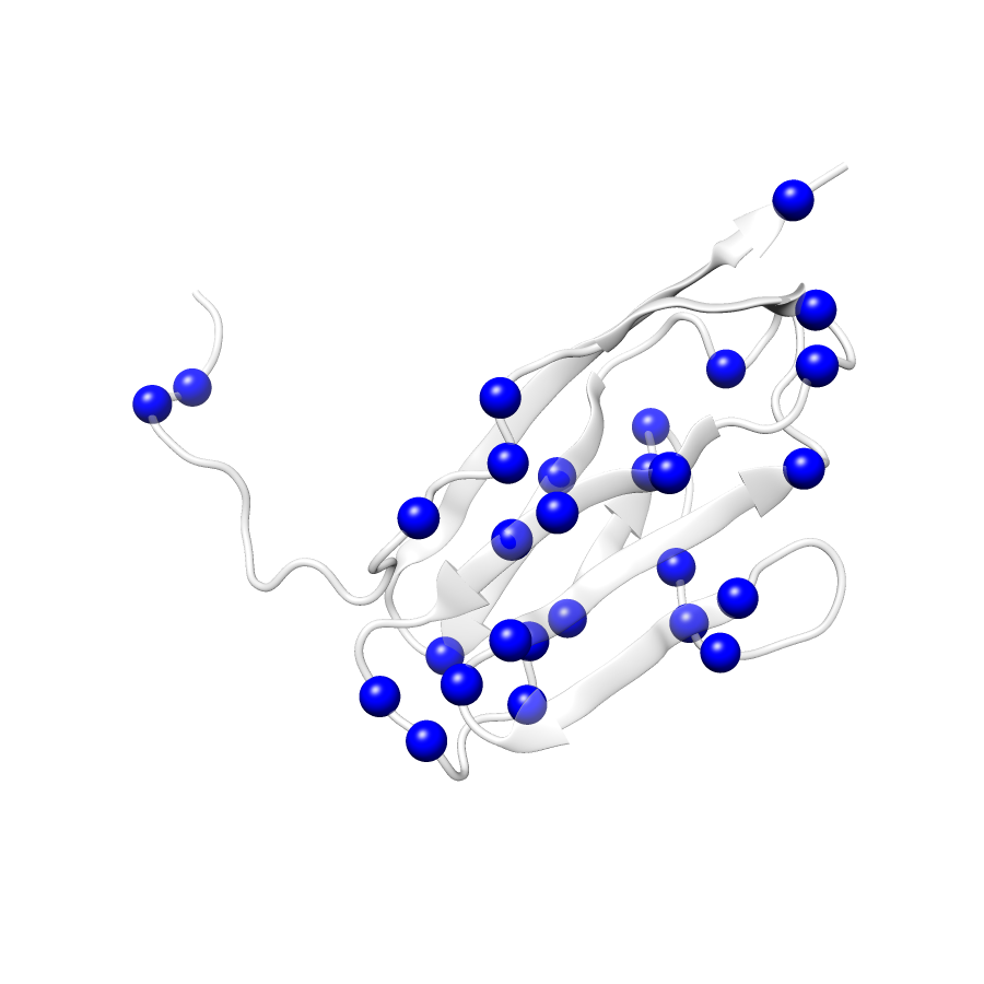
| Number Of Residues | 95 |
| Number Of SNVs | 29 |
| Number Of Permutations | 13063 |
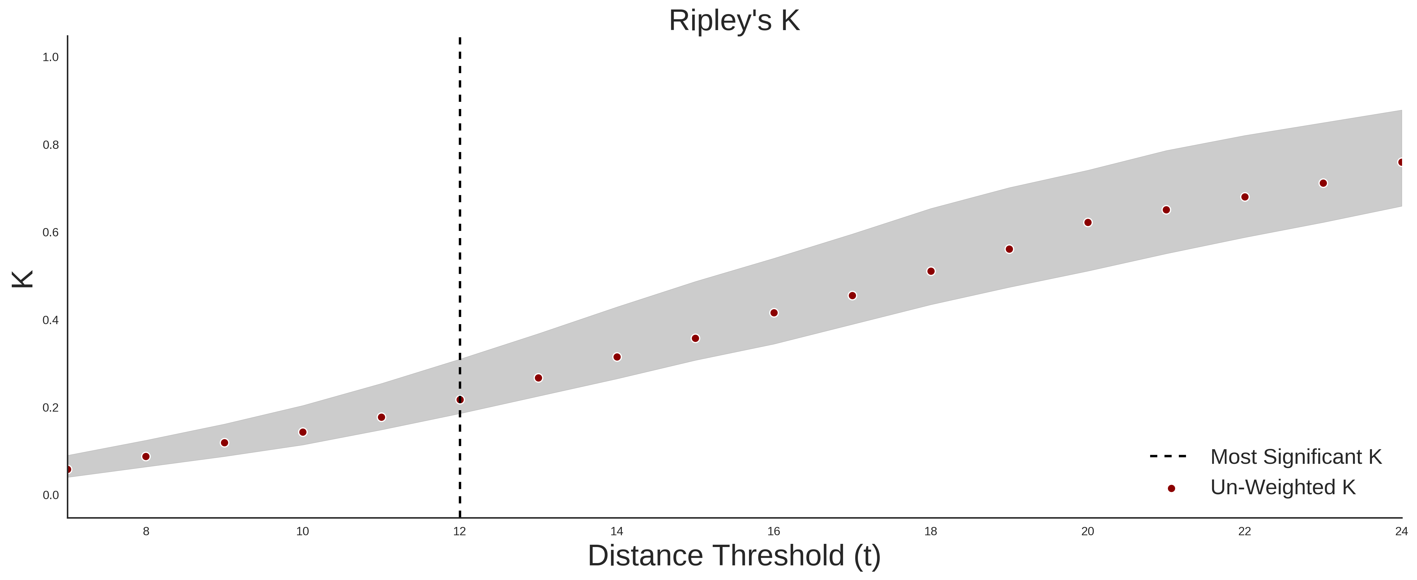
| Optimal Distance Threshold | 7.0 |
| K Statistic | 0.054 |
| p-value | 0.847 |
ClinVar
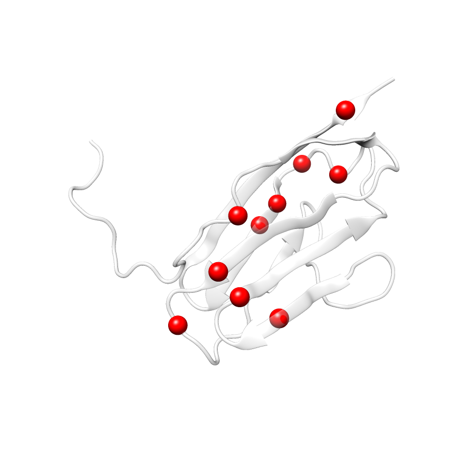
| Number Of Residues | 95 |
| Number Of SNVs | 9 |
| Number Of Permutations | 3750 |
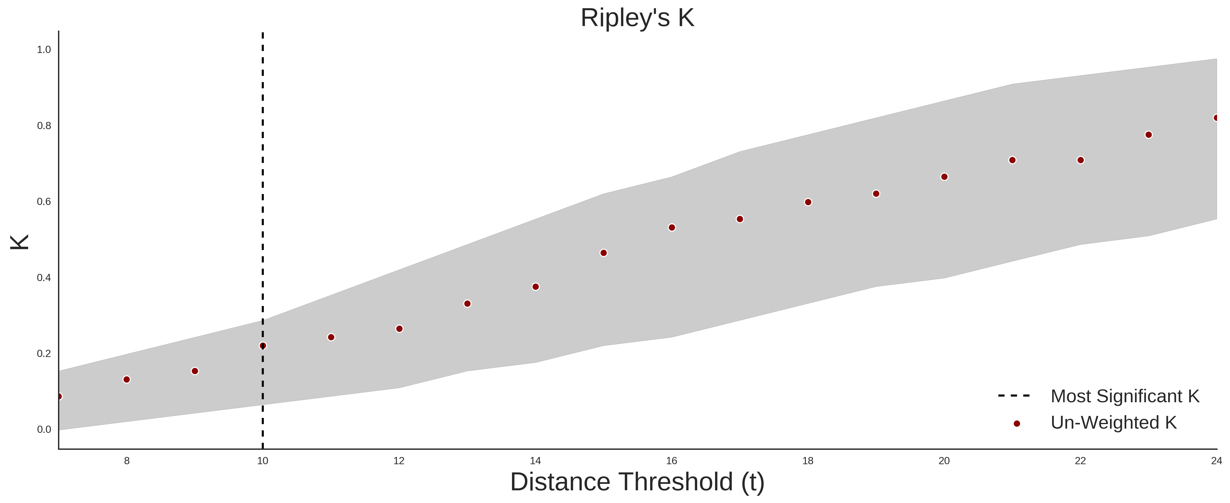
| Optimal Distance Threshold | 10.0 |
| K Statistic | 0.222 |
| p-value | 0.504 |
Ripley’s K Analysis Plots
ExACClinVar
Variant Set Comparisons
ClinVar vs. ExAC
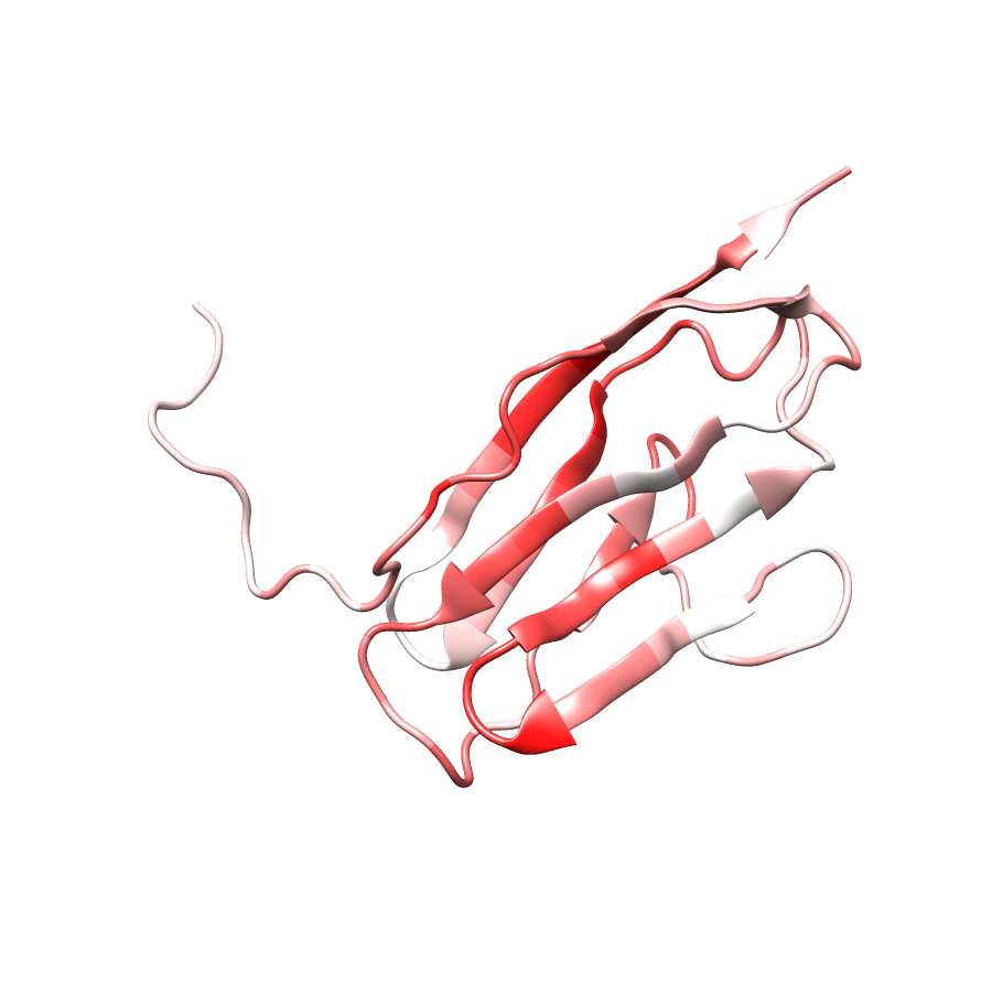
| Number Of ExAC SNVs | 29 |
| Number Of ClinVar SNVs | 8 |
| Optimal Distance Threshold | 5.0 |
| K Statistic | -0.02 |
| p-value | 0.913 |