General Structure Information
| PDB ID | 2eyi |
| HGNC Gene Label(s) | ACTN1 |
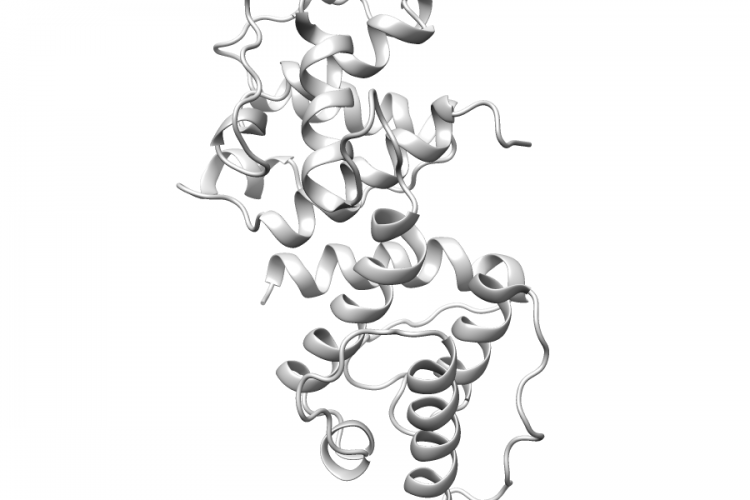
| Structure Name | crystal structure of the actin-binding domain of human alpha-actinin 1 at 1.7 angstrom resolution |
| Resolution | 1.7Å |
| Reference | AUTH E.BORREGO-DIAZ,F.KERFF,S.H.LEE,F.FERRON,Y.LI,AUTH 2 R.DOMINGUEZTITL CRYSTAL STRUCTURE OF THE ACTIN-BINDING DOMAIN OFTITL 2 ALPHA-ACTININ 1: EVALUATING TWO COMPETINGTITL 3 ACTIN-BINDING MODELS.REF J.STRUCT.BIOL. V. 155 230 2006REFN ISSN 1047-8477PMID 16698282DOI 10.1016/J.JSB.2006.01.013 |
Variant Set Distributions
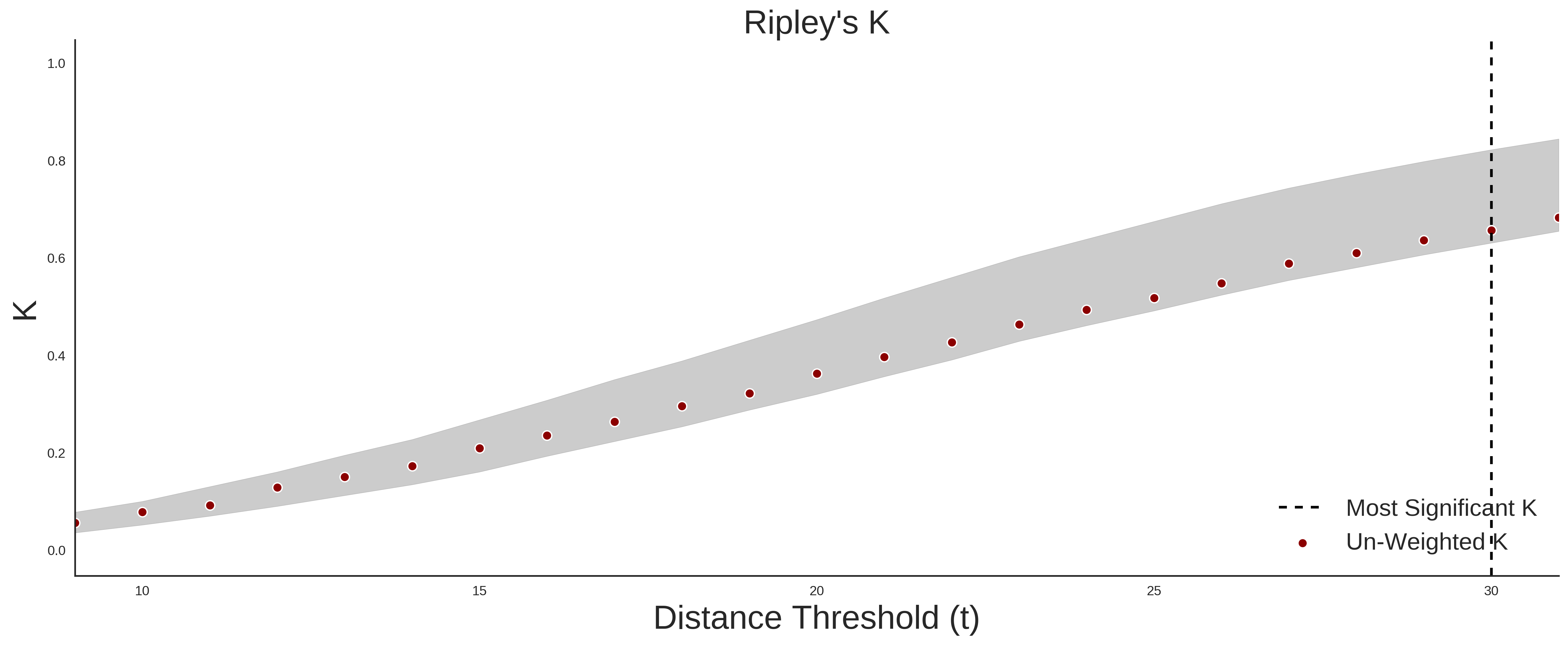
ExAC Variants
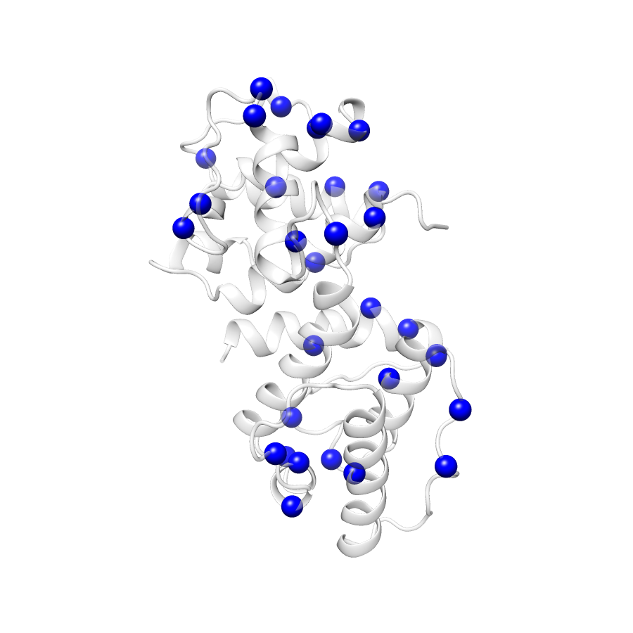
| Number Of Residues | 224 |
| Number Of SNVs | 32 |
| Number Of Permutations | 16594 |
| Optimal Distance Threshold | 19.0 |
| K Statistic | 0.323 |
| p-value | 0.681 |
ClinVar
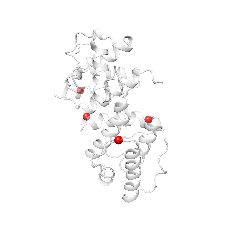
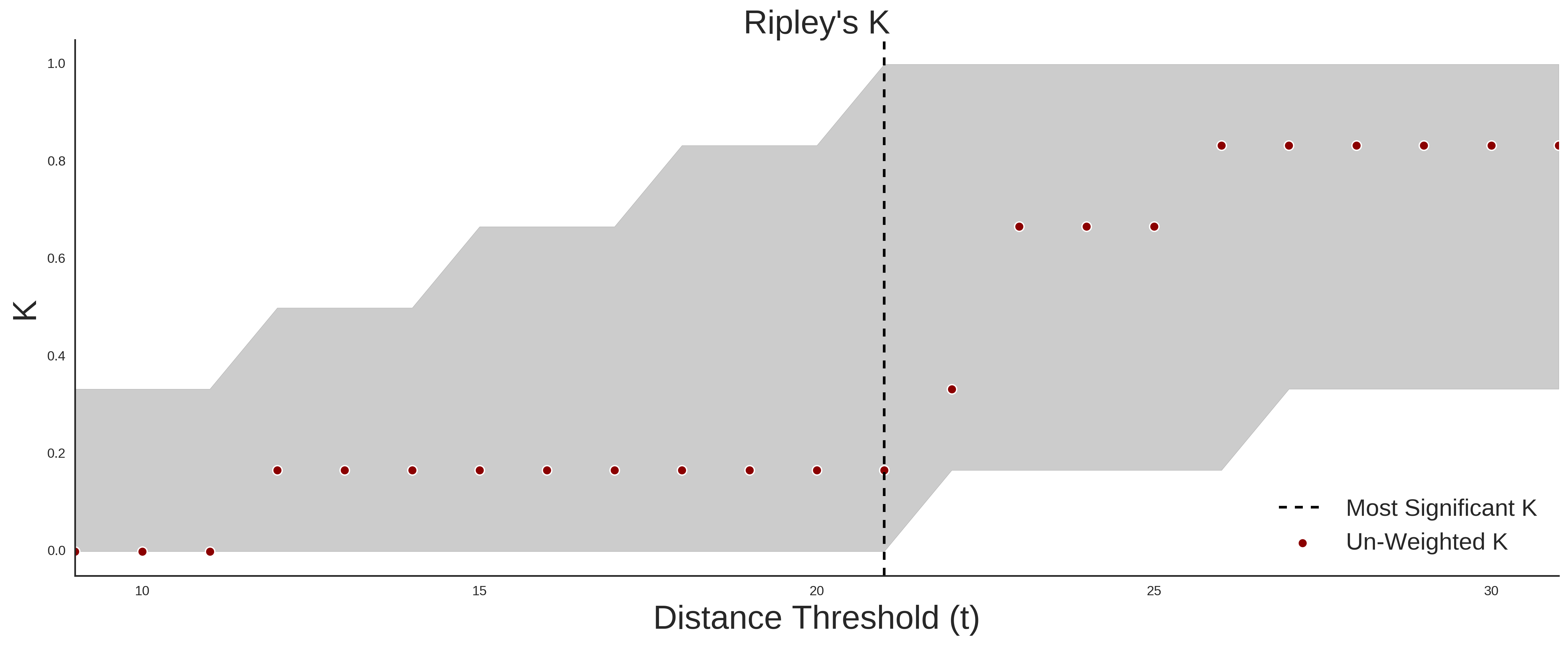
| Number Of Residues | 224 |
| Number Of SNVs | 4 |
| Number Of Permutations | 844 |
| Optimal Distance Threshold | 19.0 |
| K Statistic | 0.167 |
| p-value | 0.697 |
Ripley’s K Analysis Plots
ExACClinVar
Variant Set Comparisons
ClinVar vs. ExAC
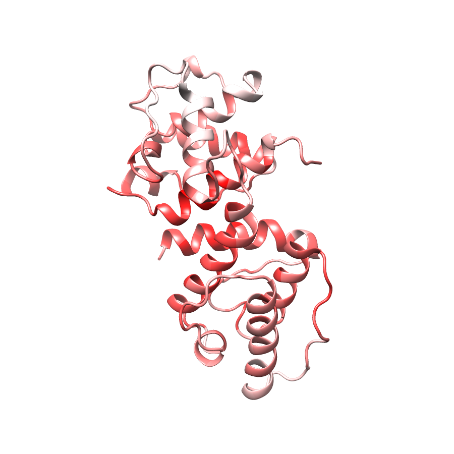
| Number Of ExAC SNVs | 32 |
| Number Of ClinVar SNVs | 4 |
| Optimal Distance Threshold | 11.0 |
| K Statistic | -0.091 |
| p-value | 0.863 |
Pathogenic Proximity Analysis
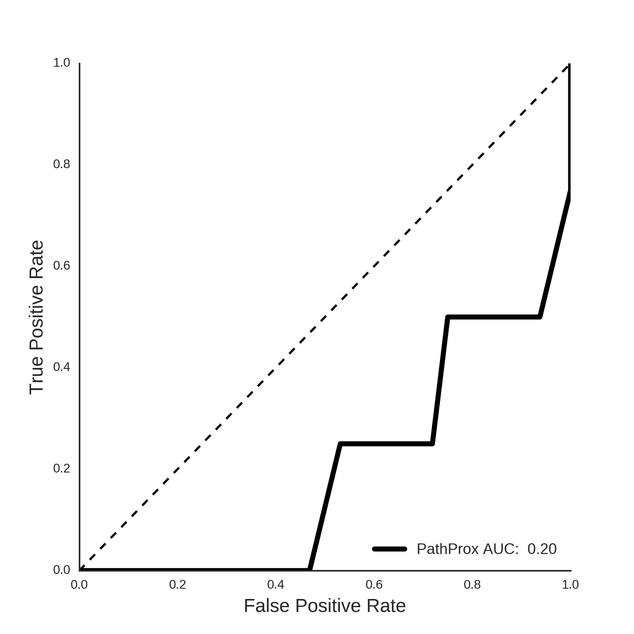
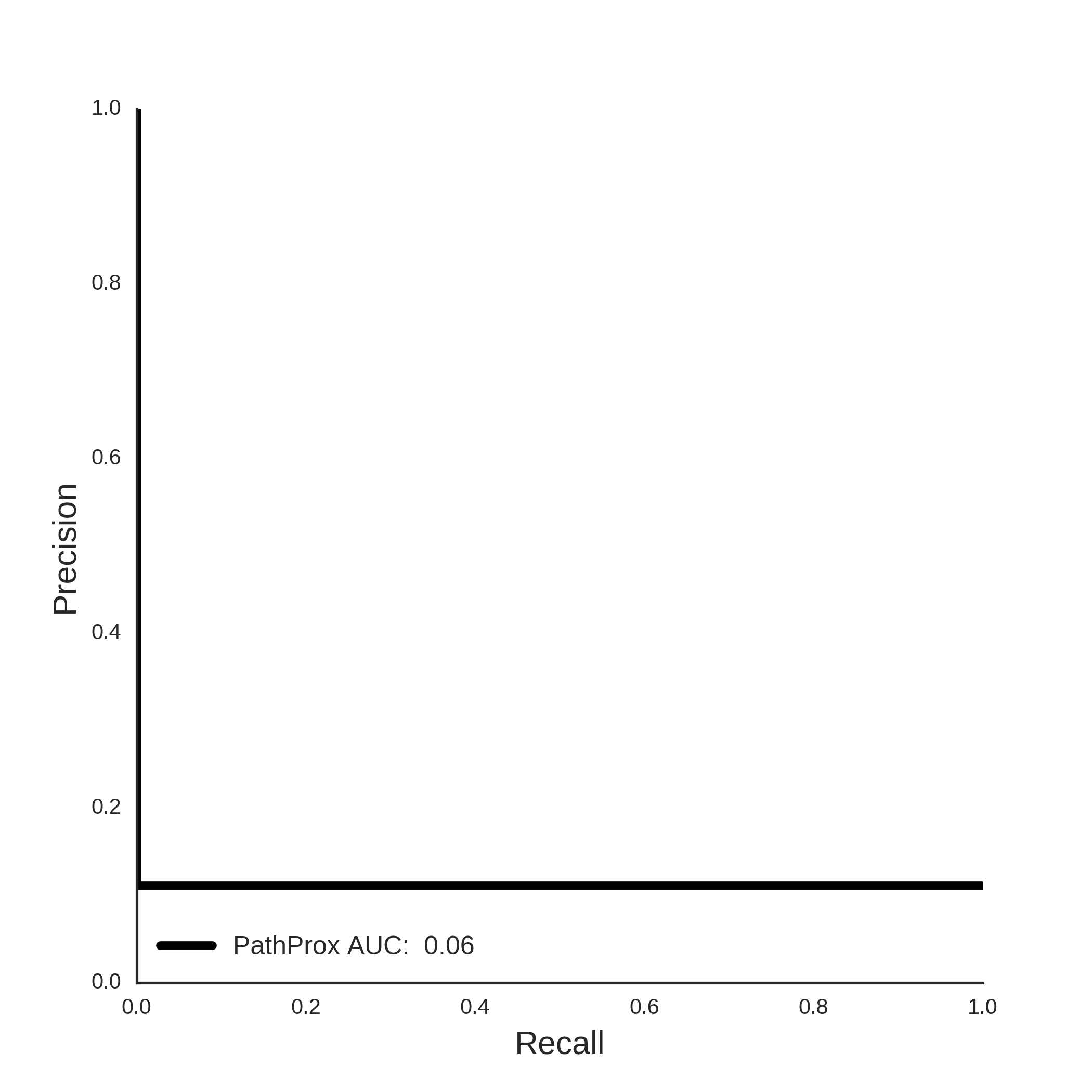
ClinVar PathProx Analysis