General Structure Information
| PDB ID | 2bcd |
| HGNC Gene Label(s) | PPP1CC |
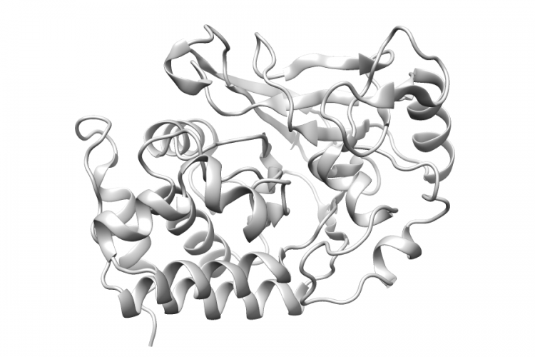
| Structure Name | x-ray crystal structure of protein phosphatase-1 with the marine toxin motuporin bound |
| Resolution | 2.1Å |
| Reference | AUTH J.T.MAYNES,H.A.LUU,M.M.CHERNEY,R.J.ANDERSEN,D.WILLIAMS,AUTH 2 C.F.HOLMES,M.N.JAMESTITL CRYSTAL STRUCTURES OF PROTEIN PHOSPHATASE-1 BOUND TOTITL 2 MOTUPORIN AND DIHYDROMICROCYSTIN-LA: ELUCIDATION OF THETITL 3 MECHANISM OF ENZYME INHIBITION BY CYANOBACTERIAL TOXINS.REF J.MOL.BIOL. V. 356 111 2006REFN ISSN 0022-2836PMID 16343532DOI 10.1016/J.JMB.2005.11.019 |
Variant Set Distributions
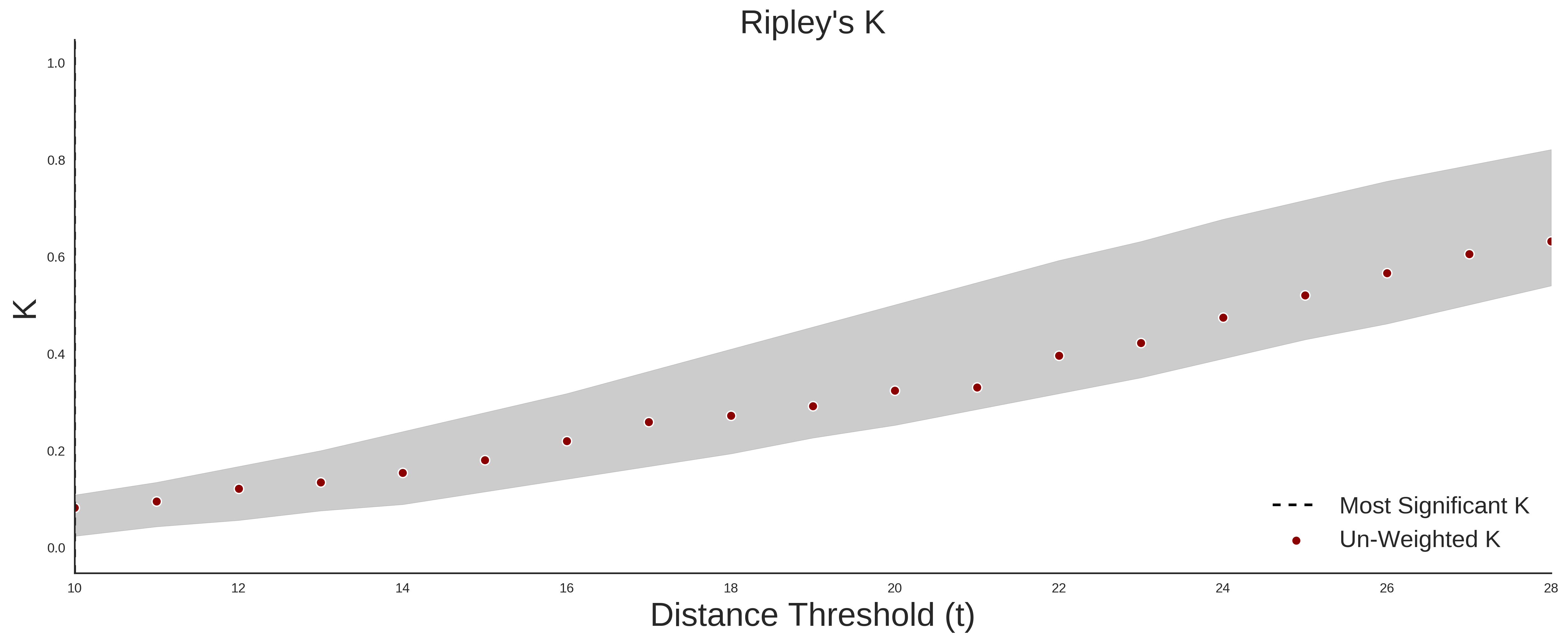
ExAC Variants
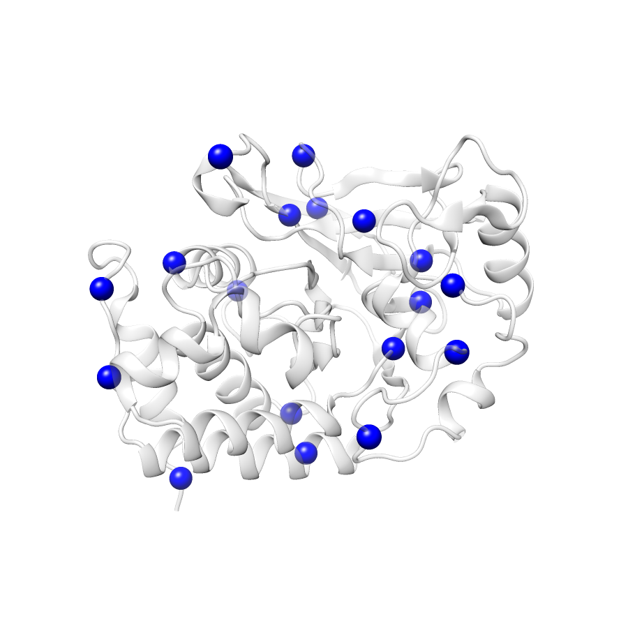
| Number Of Residues | 293 |
| Number Of SNVs | 18 |
| Number Of Permutations | 6963 |
| Optimal Distance Threshold | 9.0 |
| K Statistic | 0.065 |
| p-value | 0.67 |
COSMIC
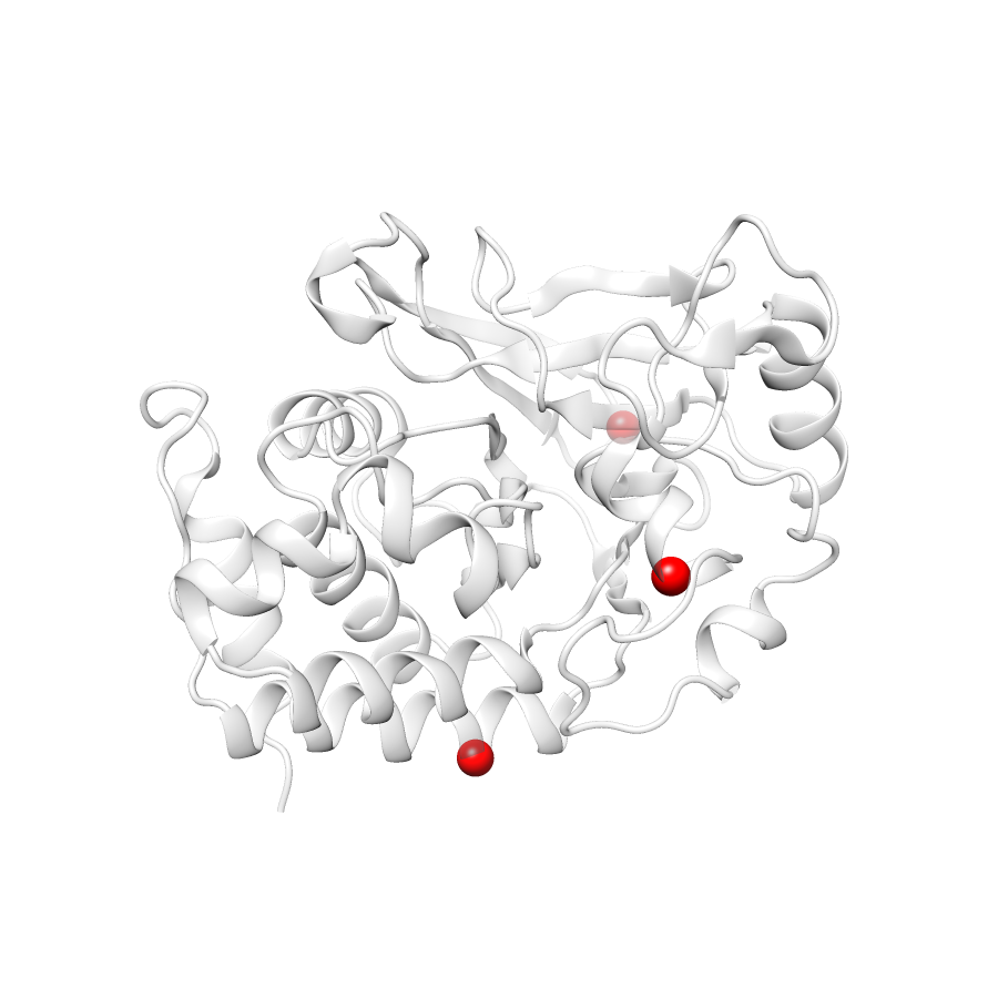
| Number Of Residues | 293 |
| Number Of SNVs | 3 |
| Number Of Permutations | 159 |
| Optimal Distance Threshold | 27.0 |
| K Statistic | 0.333 |
| p-value | 0.713 |
Ripley’s K Analysis Plots
ExACCOSMIC
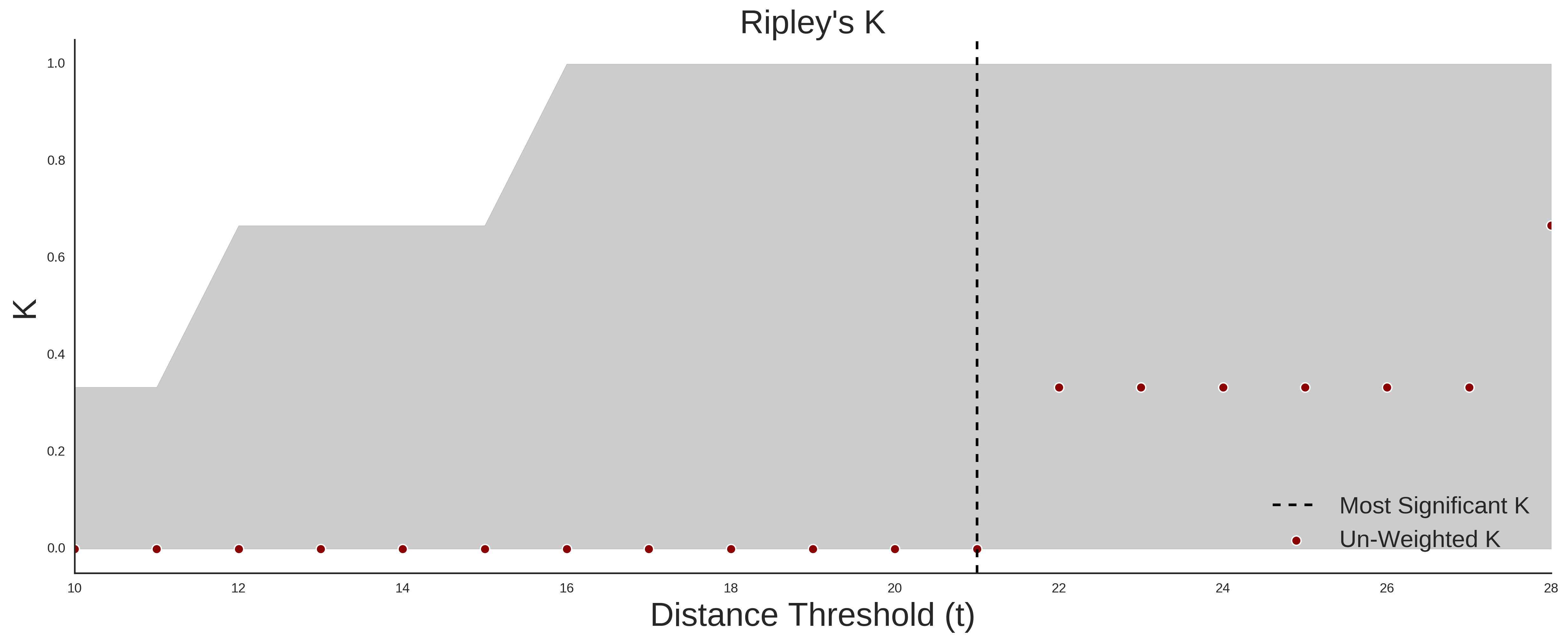
Variant Set Comparisons
Cosmic vs. ExAC
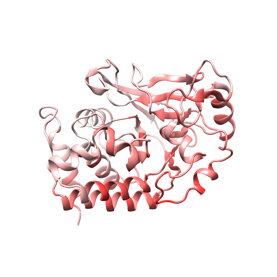
Pathogenic Proximity Analysis
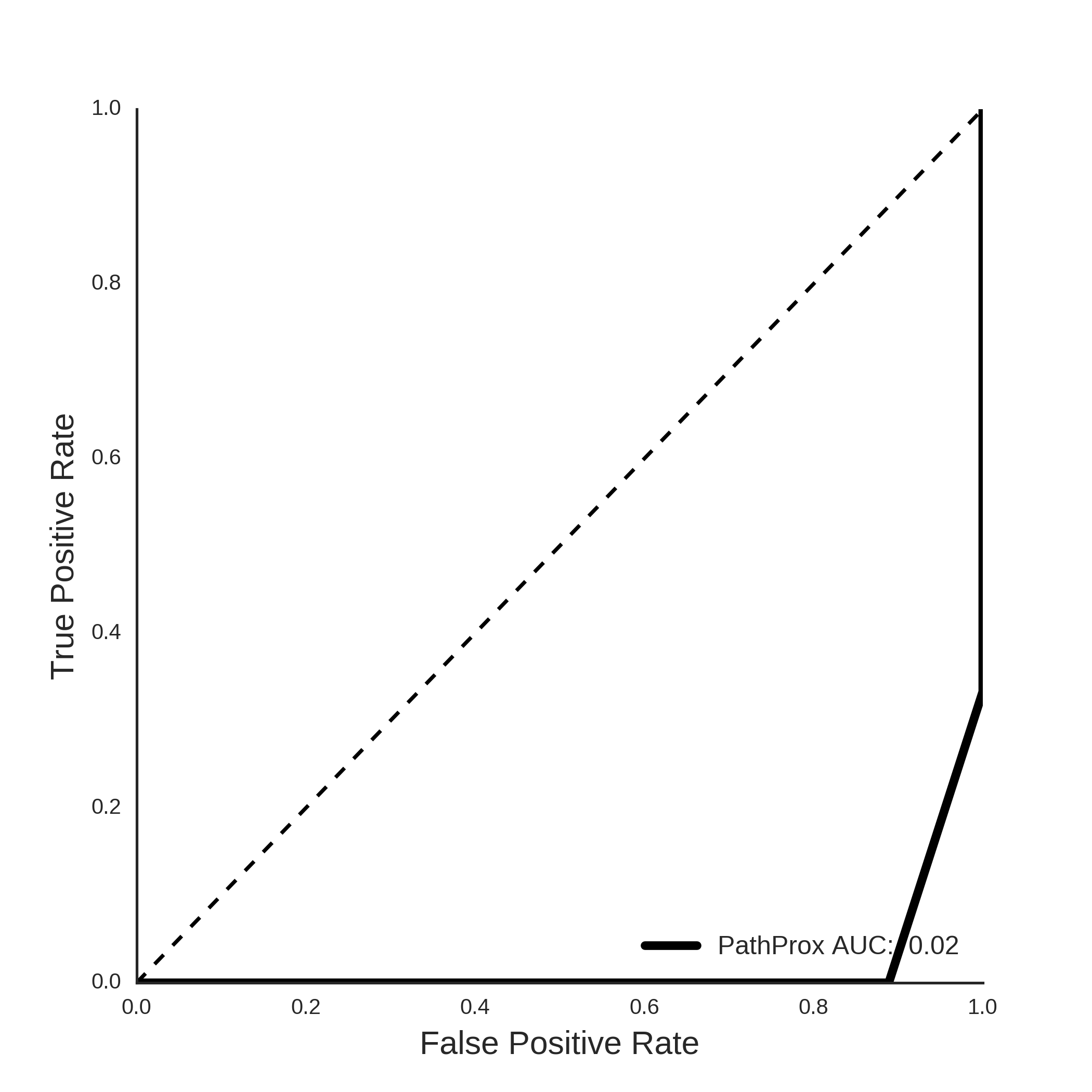
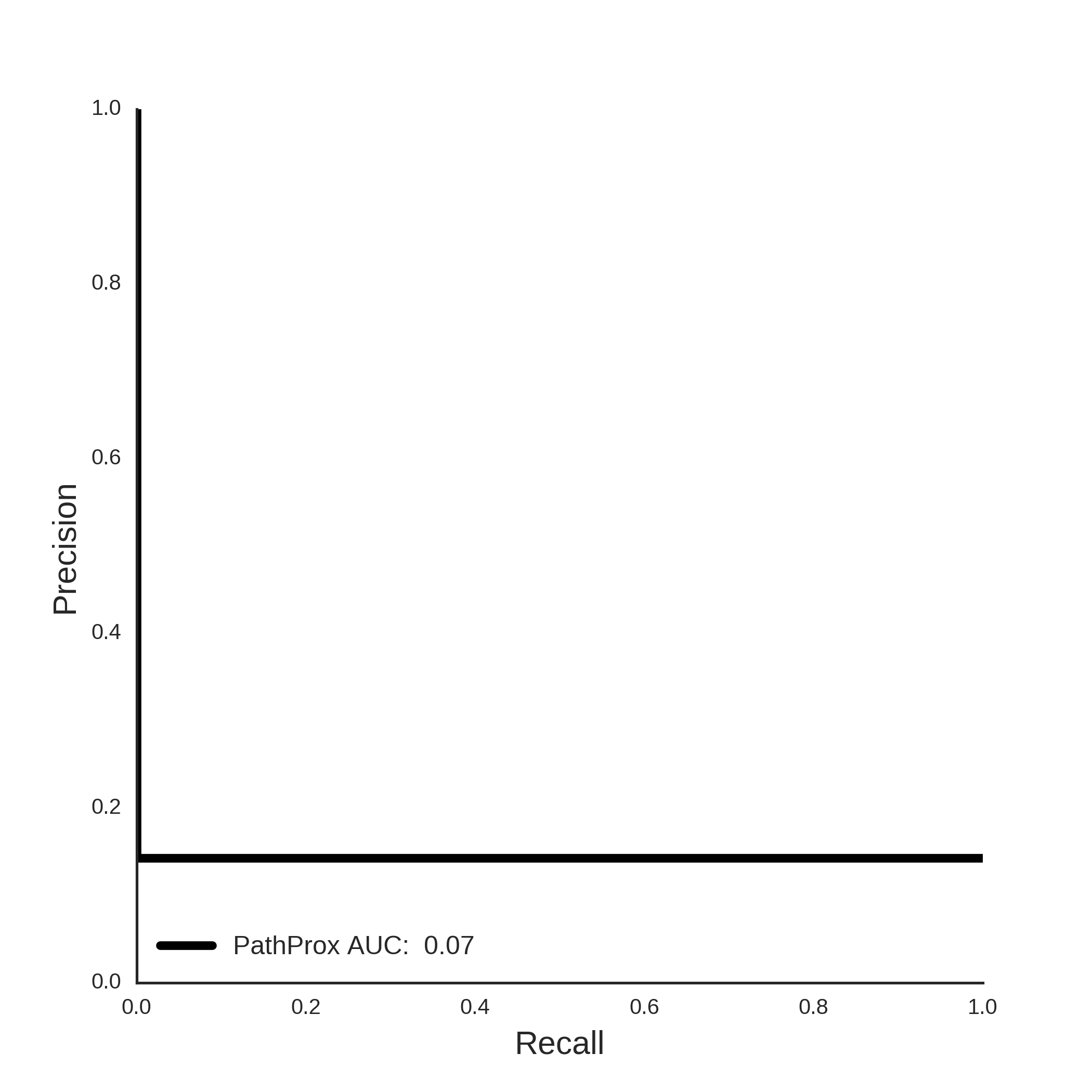
COSMIC PathProx Analysis