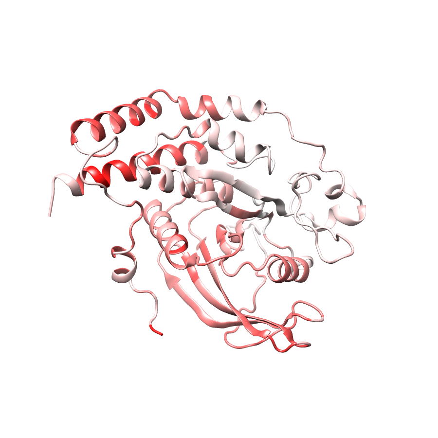
General Structure Information
| PDB ID | 1wch |
| HGNC Gene Label(s) | PTPN13 |
| Structure Name | crystal structure of ptpl1 human tyrosine phosphatase mutated in colorectal cancer – evidence for a second phosphotyrosine substrate recognition pocket |
| Resolution | 1.85Å |
| Reference | AUTH F.VILLA,M.DEAK,G.B.BLOOMBERG,D.R.ALESSI,AUTH 2 D.M.F.VAN AALTENTITL CRYSTAL STRUCTURE OF PTPL1/FAP-1 HUMAN TYROSINETITL 2 PHOSPHATASE MUTATED IN COLORECTAL CANCER:TITL 3 EVEIDENCE FOR A SECOND PHOSPHOTYROSINE SUBSTRATETITL 4 RECOGNITION POCKETREF J.BIOL.CHEM. V. 280 8180 2005REFN ISSN 0021-9258PMID 15611135DOI 10.1074/JBC.M412211200 |
Variant Set Distributions
ExAC Variants
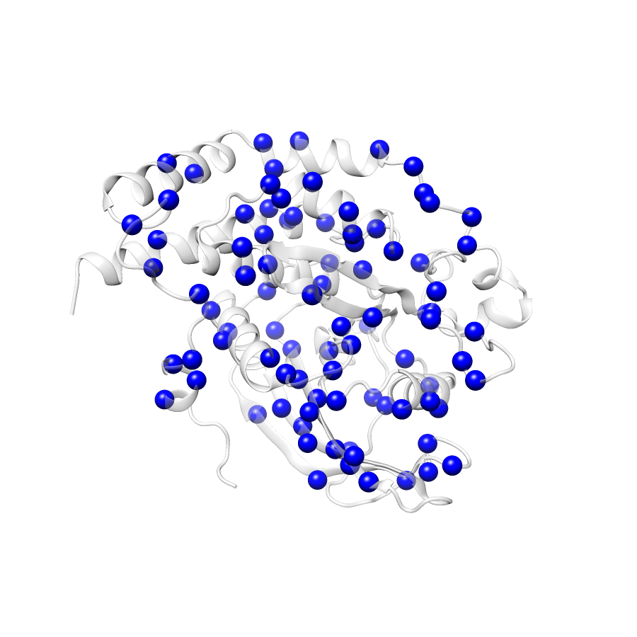
| Number Of Residues | 308 |
| Number Of SNVs | 88 |
| Number Of Permutations | 16296 |
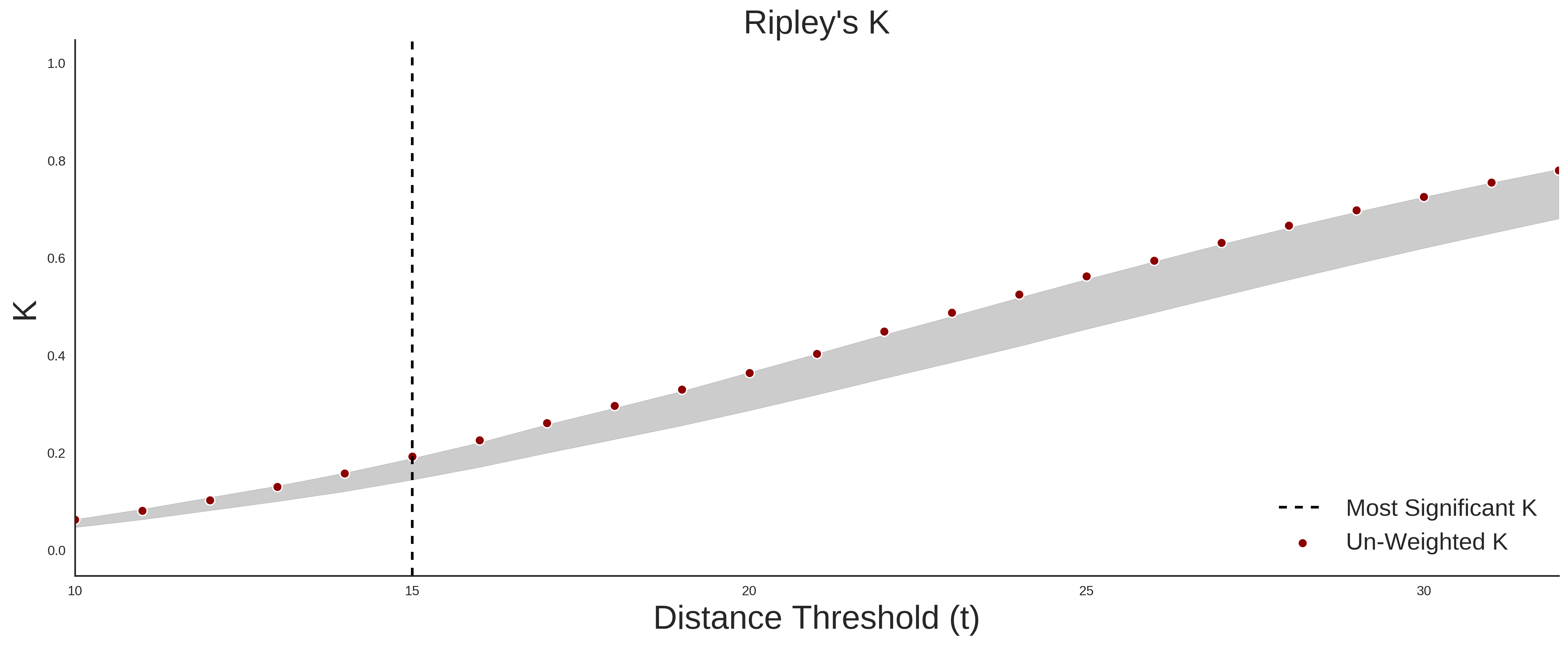
| Optimal Distance Threshold | 15.0 |
| K Statistic | 0.189 |
| p-value | 0.111 |
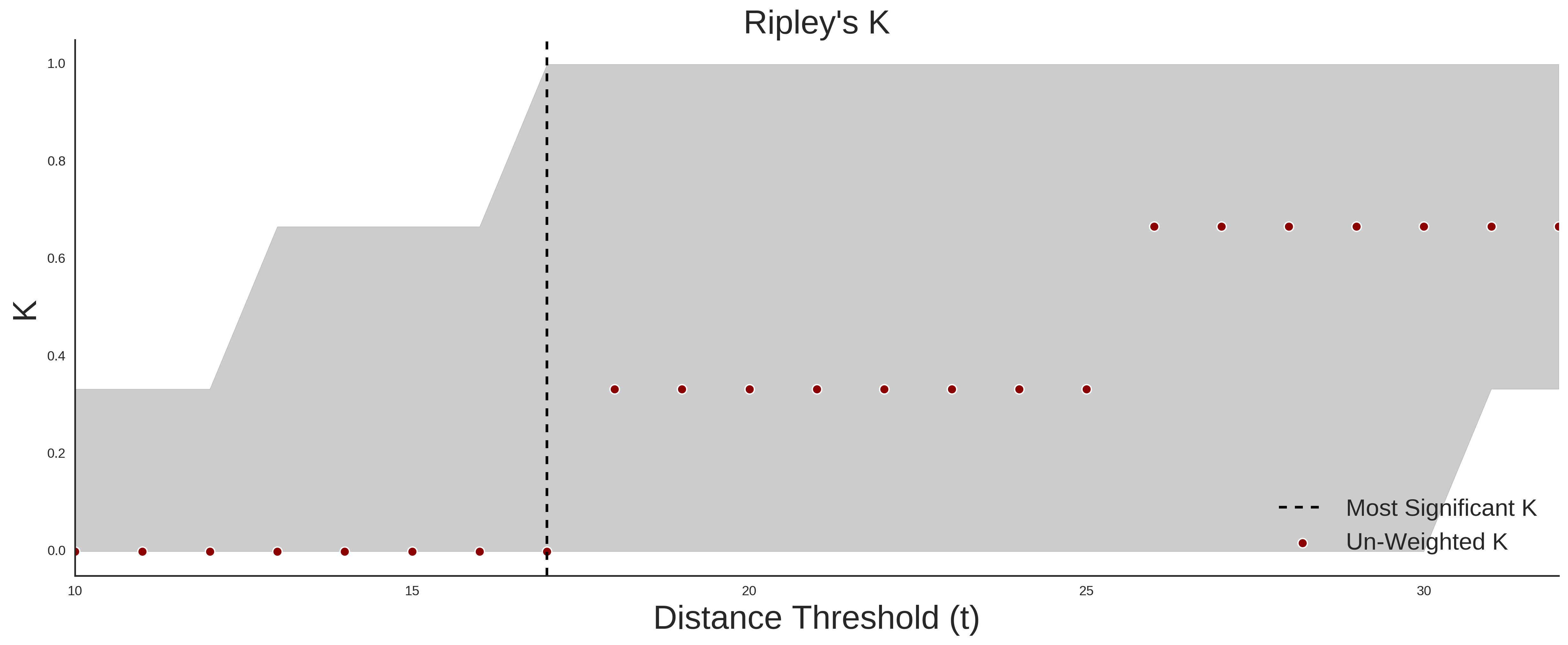
COSMIC
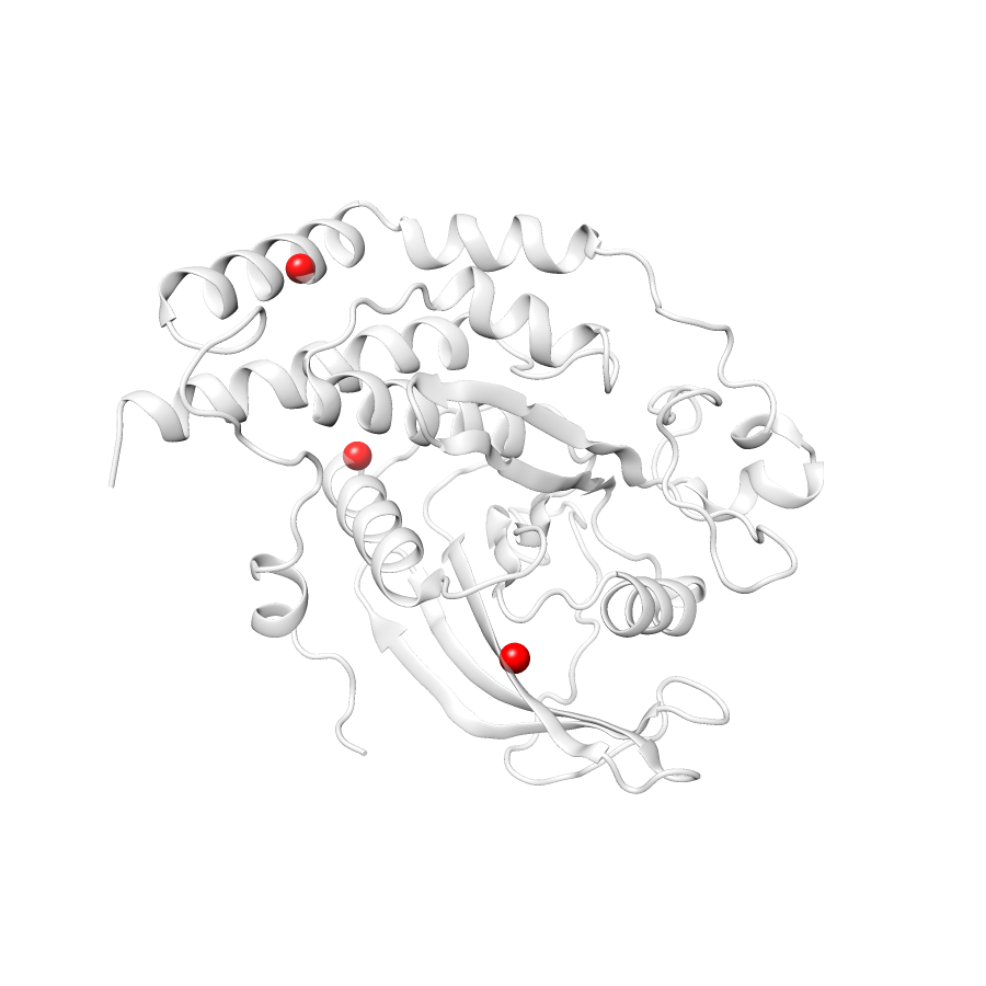
| Number Of Residues | 308 |
| Number Of SNVs | 3 |
| Number Of Permutations | 18 |
| Optimal Distance Threshold | 18.0 |
| K Statistic | 0.333 |
| p-value | 1.0 |
Ripley’s K Analysis Plots
ExACCOSMIC
Variant Set Comparisons
Cosmic vs. ExAC